
Dimitri Aubert


The Digital Twin of Scattering Light
This artwork illustrates the research by Mohammadrahim Kazemzadeh, Massimo De Vittorio, Ferruccio Pisanello, and co-workers, who developed a physics-informed neural network capable of creating a digital twin of light traveling through complex, scattering materials.
The concept merges physics and artificial intelligence to reveal how light propagates in turbid media, transforming raw data into an interpretable model of illumination and structure. (Istituto Italiano di Tecnologia).
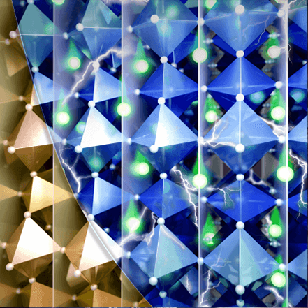
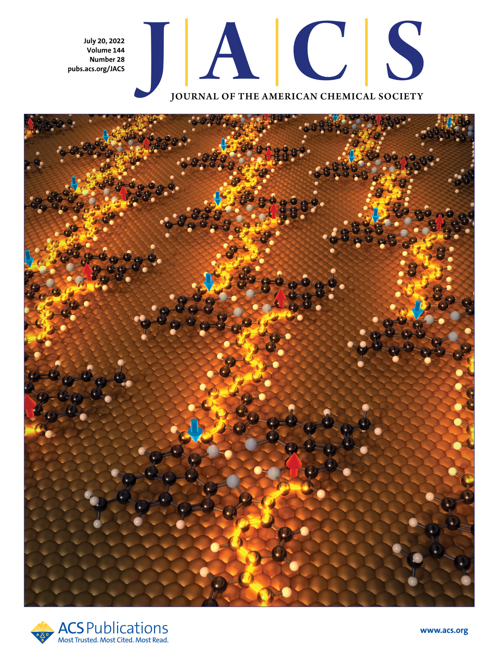
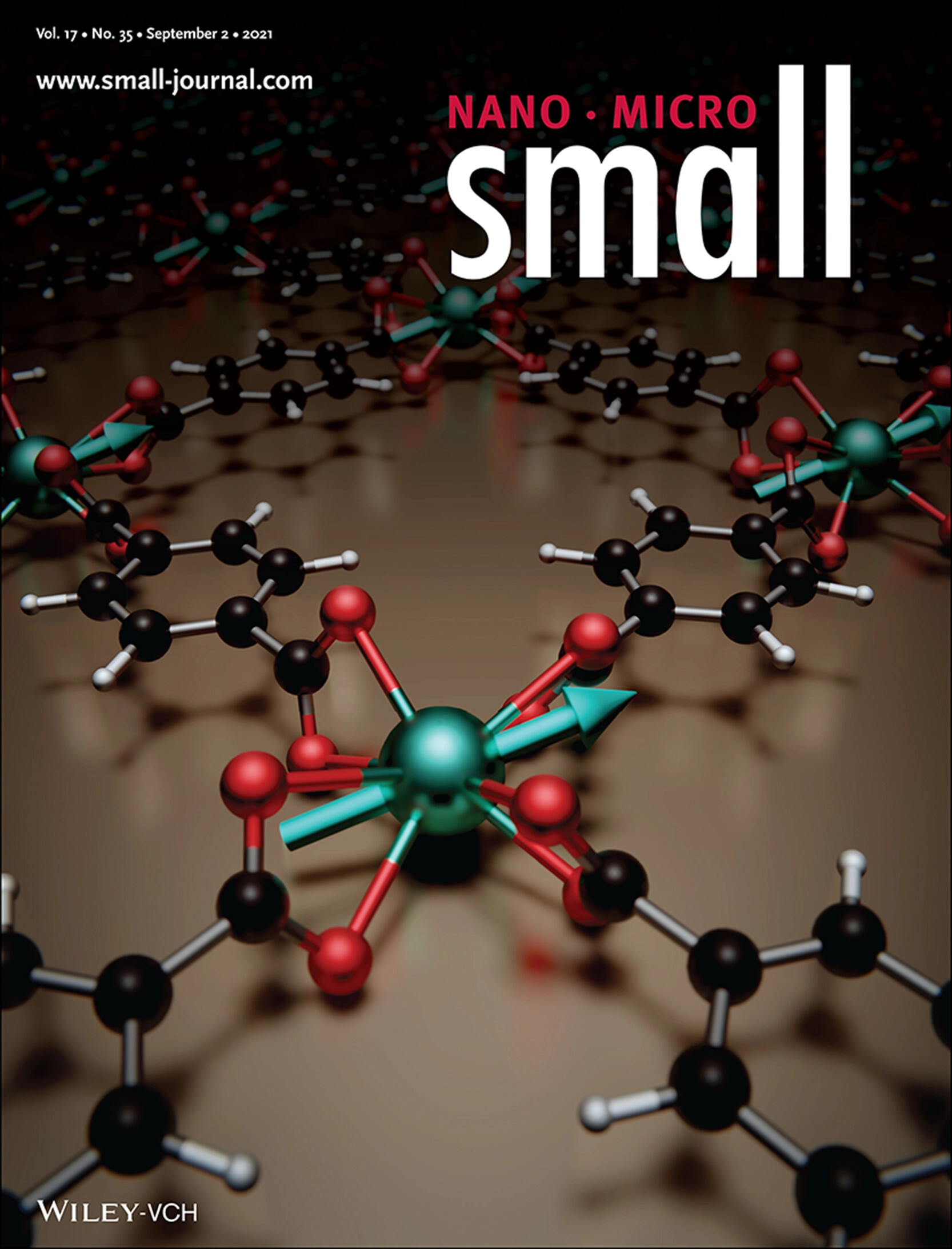
Engineering a Spin-Orbit Bandgap in Graphene-Tellurium Heterostructures
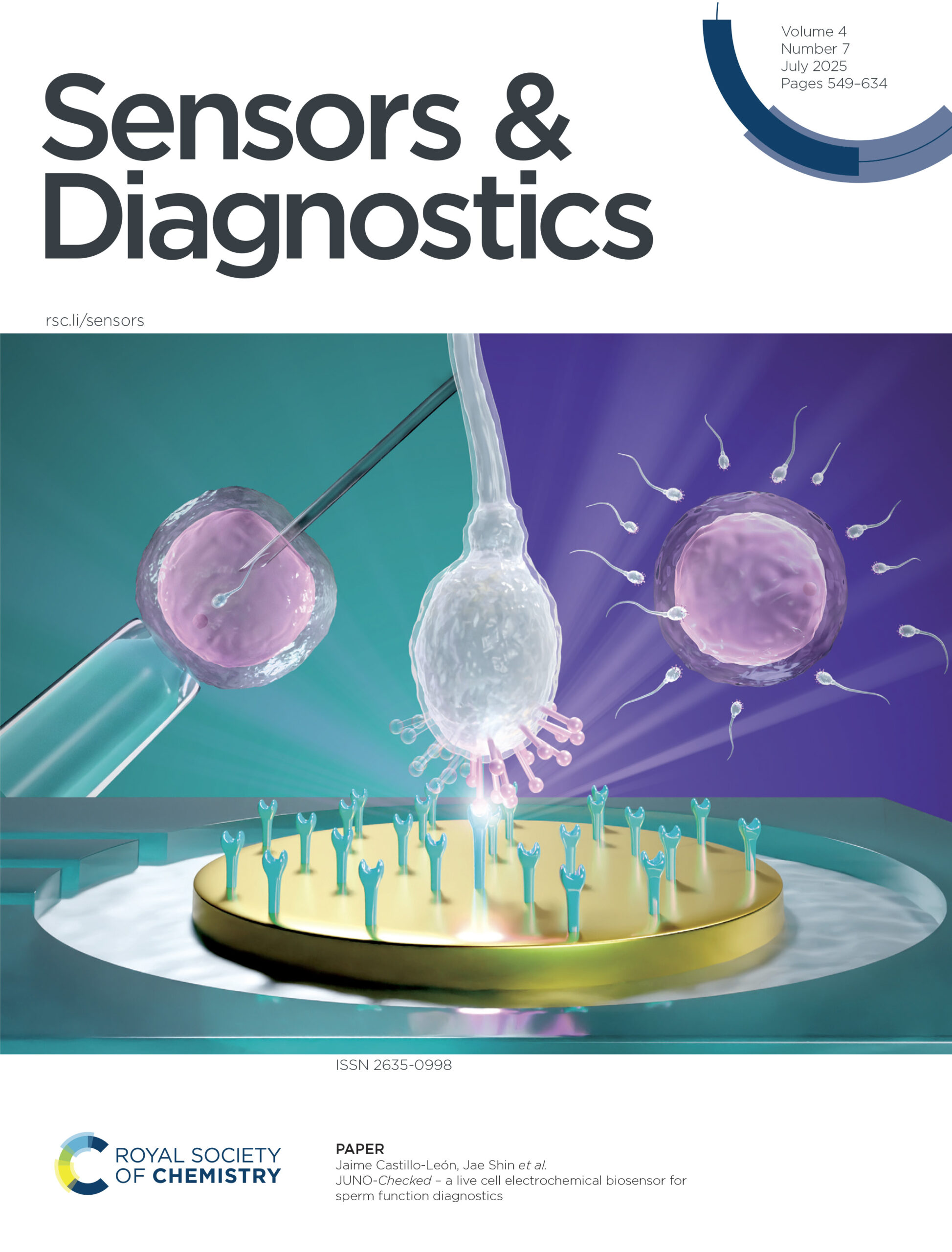
This journal cover artwork illustrates the research by Jaime Castillo-León, Jae Shin, and their team at Spermosens AB, who developed JUNO-Checked — an innovative live-cell electrochemical biosensor designed to evaluate sperm function with exceptional precision. Inspired by the molecular mechanisms of fertilization, the device uses immobilized JUNO proteins, naturally found on oocytes, to assess how effectively sperm can bind to the egg surface. This interaction generates a quantitative metric called the JUNOScore, offering a new dimension of information about sperm functionality — one that traditional microscopic semen analyses cannot capture. By revealing these deeper functional insights, JUNO-Checked can help clinicians tailor personalized assisted reproduction treatments, improving diagnostic precision in male fertility assessment.
Catalyst-Free Graphene Growth
This artwork represents the research by Mustapha Jouiad, Ahmed Kotbi, Michael Lejeune, and co-workers, who developed an eco-friendly, catalyst-free method for growing graphene directly on diverse substrates.
Using ethylene as the only carbon source, their process forms few-layer graphene films with excellent conductivity, without complex transfers or toxic catalysts.
The image highlights the elegance of sustainable materials science: clean chemistry, simple methods, and powerful results.
Label-Like Power: Flexible Organic Thermoelectric Generators
This artwork illustrates the research by Nathan James Pataki, Mario Caironi, and co-workers (Istituto Italiano di Tecnologia), who developed ultra-thin, label-like thermoelectric generators using aligned organic polymers. Through precise inkjet doping, these flexible films can convert tiny temperature differences into electricity, like smart energy stickers. The image highlights the beauty of soft, printable electronics shaping a more sustainable future.
Engineering a Spin-Orbit Bandgap in Graphene-Tellurium Heterostructures
This artwork illustrates the research by Mohammadrahim Kazemzadeh, Massimo De Vittorio, Ferruccio Pisanello, and co-workers, who developed a physics-informed neural network capable of creating a digital twin of light traveling through complex, scattering materials.
The concept merges physics and artificial intelligence to reveal how light propagates in turbid media, transforming raw data into an interpretable model of illumination and structure. (Istituto Italiano di Tecnologia).
Mechanochemically activated Biginelli reaction
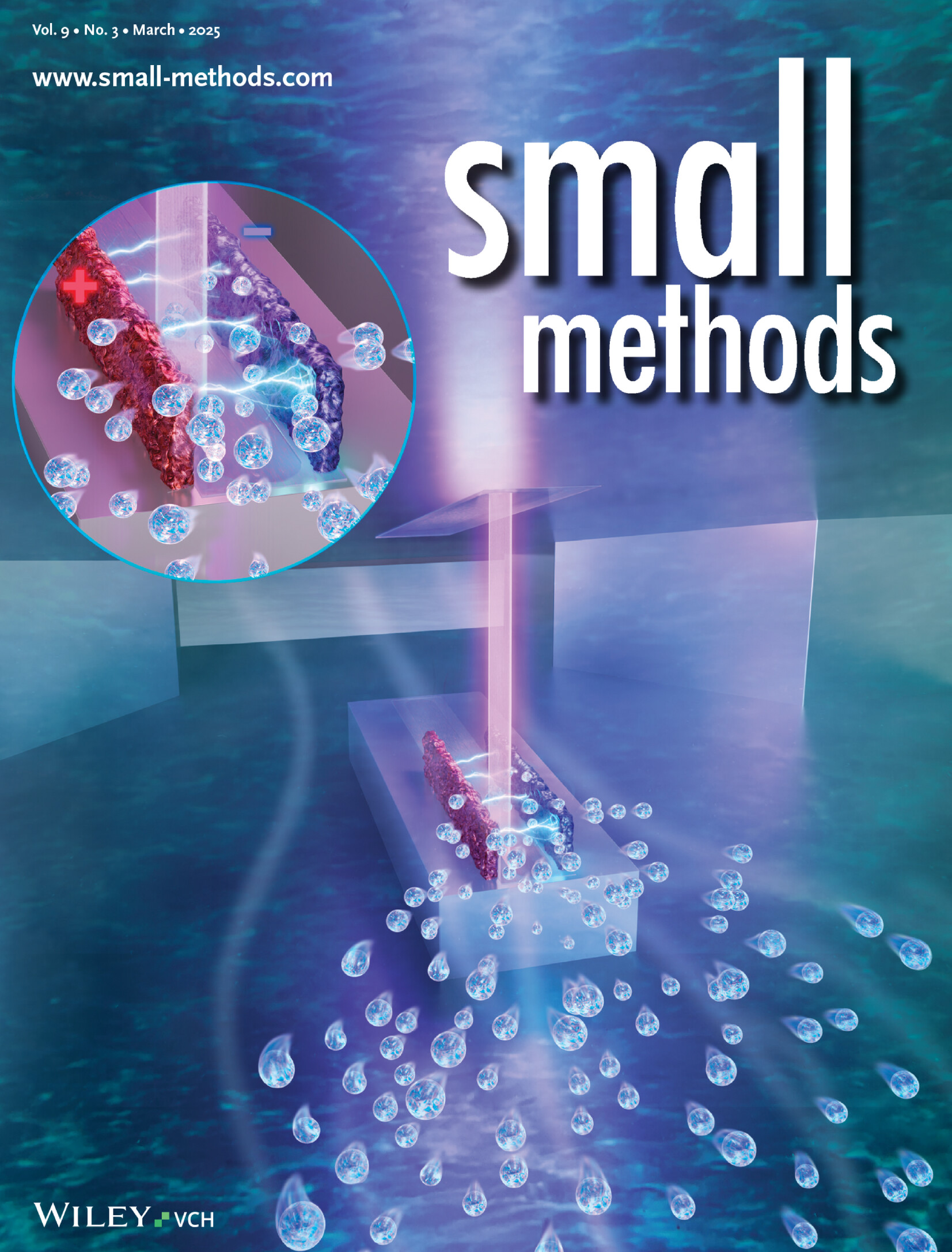
This image captures the interaction between a beam of electron radiation and an electrochemical system. At the core, two electrodes symbolize the experimental setup, while a circular inset zooms in on the heart of the system: a uniquely designed electrochemical cell with a central island.
Streamlines trace the liquid flow, which plays a crucial role in directing the system’s dynamics. Notice the tiny gas bubbles forming—these are the result of electrochemical reactions—and thanks to the clever cell design, they’re swept away efficiently by the circulating liquid.
From geometry to flow physics, this illustration reflects the interplay of design and function explored in work of Fontana and co-workers (Istituto Italiano di Tecnologia).
Electrochemical Random Access Memory
This illustration depicts an Electrochemical Random Access Memory (ECRAM), a technology that operates by moving mobile defects—such as protons, lithium ions, or oxygen vacancies—between the top (reservoir) and bottom (channel) electrodes, connected by a solid electrolyte (light blue). Through reversible redox reactions, these mobile defects tune the electronic conductance, enabling efficient and scalable memory solutions for computing.
Inspired by the work of A. Alec Talin and his colleagues (Sandia National Laboratories), this image represents the fundamental principles behind ECRAM’s potential for next-generation electronics.
Compositional site disorder
This illustration represents the role of compositional site disorder in modifying oxide semiconductors. The work of Elliot J. Fuller and colleagues (Sandia National Laboratories) demonstrates how controlled disorder can alter carrier type, enhance crystallinity, and tune spin transitions, enabling applications in electrothermal thresholding devices like radio frequency limiters.
A visualization of how structural manipulation at the atomic level can lead to new functionalities in advanced materials.
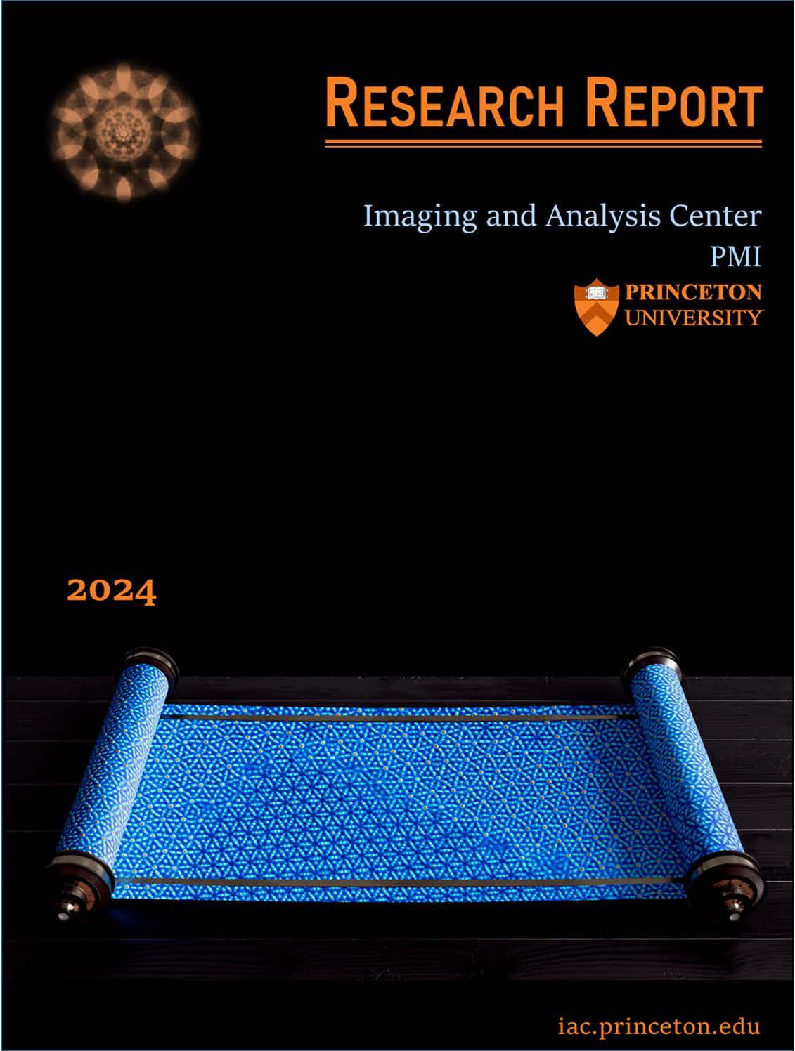
Research Report
A scientific visualization developed for the 2024 Research Report, inspired by Prof. Nan Yao’s research at Princeton University.
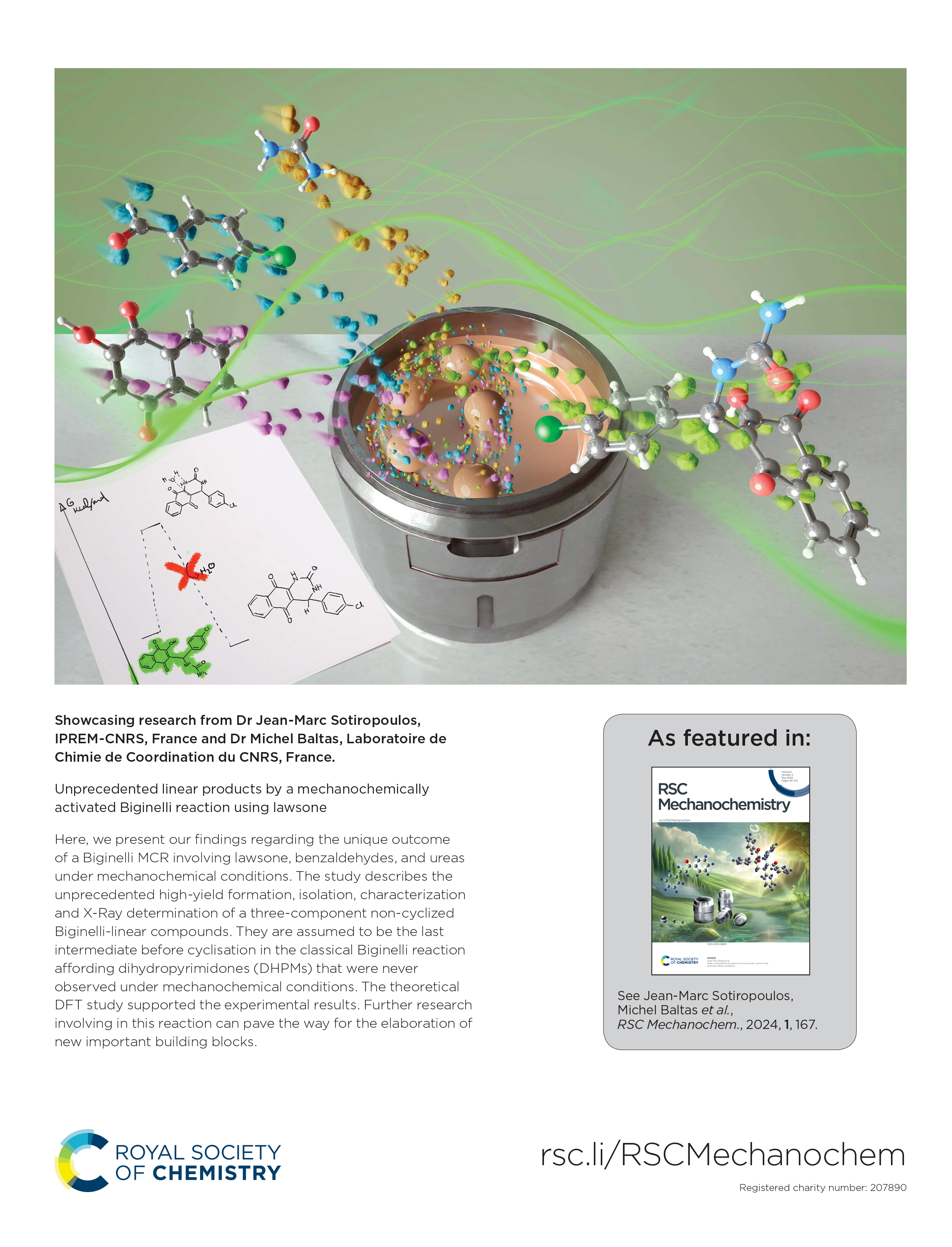
Mechanochemically activated Biginelli reaction
This illustration visualizes the outcome of a Biginelli multicomponent reaction (MCR) under mechanochemical conditions. The study, led by Christina Koumpoura, PhD, Marc Sotiropoulos, Michel Baltas, and colleagues (CNRS-IPREM & CNRS-LCC), reveals the high-yield formation and characterization of unprecedented non-cyclized Biginelli-linear compounds, believed to be the last intermediate before cyclization into dihydropyrimidones (DHPMs).
Supported by DFT theoretical studies, these findings open new possibilities for designing novel building blocks in organic synthesis.
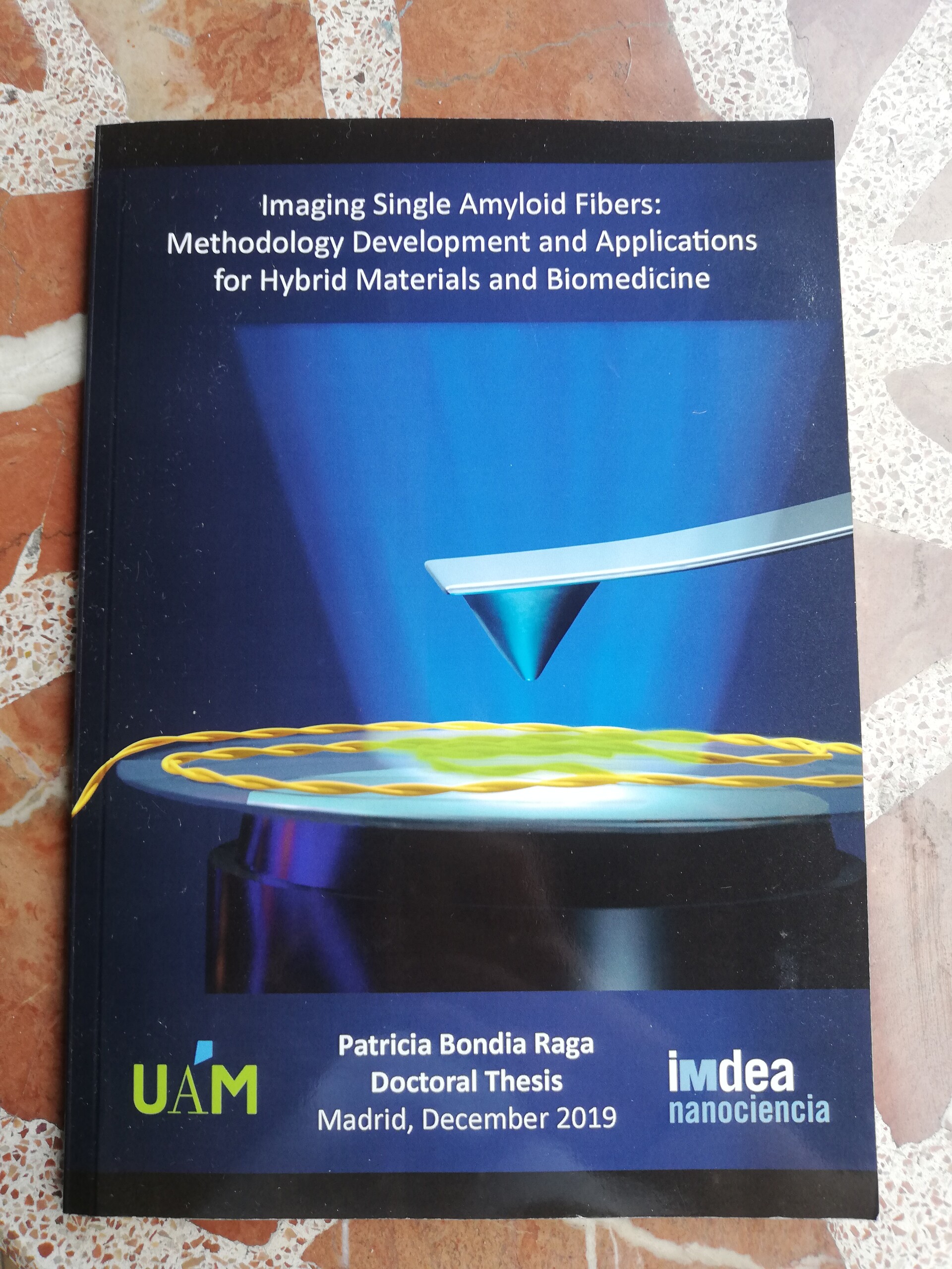
Nanoscale View of Amyloid Photodynamic Damage
The image illustrates the impact of a light-activated molecule (ROS-ThT), effectively breaking the targeted amyloid fiber. In the context of biomedicine, this study holds crucial importance, as it explores new strategies for treating neurodegenerative diseases such as Alzheimer’s or Parkinson’s. The artwork was created for Cristina Flors’ research group, it holds particular significance as it forms the main focus of my thesis and marks the occasion of my first published cover, representing a significant milestone in my academic and illustrator journey.
This research is the result of a collaborative effort between IMDEA Nanoscience and Tokyo University.
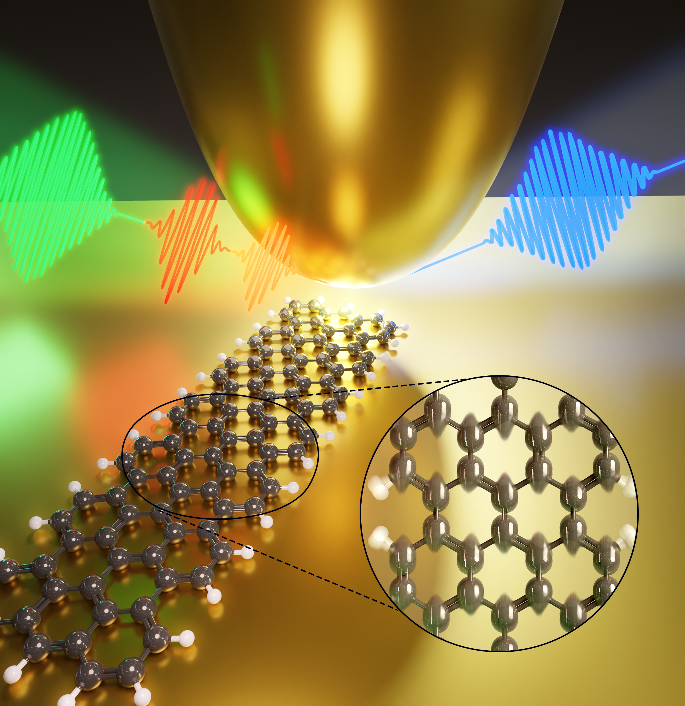
Can we watch and control the movement of atoms within a molecule?
A study led by Manish Garg, Alberto Martín and coworkers (MPI-FKF Stuttgart and UAM) has answered that question. By performing ultrafast spectroscopy within a scanning tunneling microscope, they revealed the periodic vibrations of atoms and showcased their ability to capture and finely manipulate them.
This image illustrates how the atoms within a single graphene nanoribbon (GNR) were set in motion by two delay-controlled ultrashort pulses. A third pulse, performs anti-Stokes Raman spectroscopy, tracked the vibrational features within the GNR.
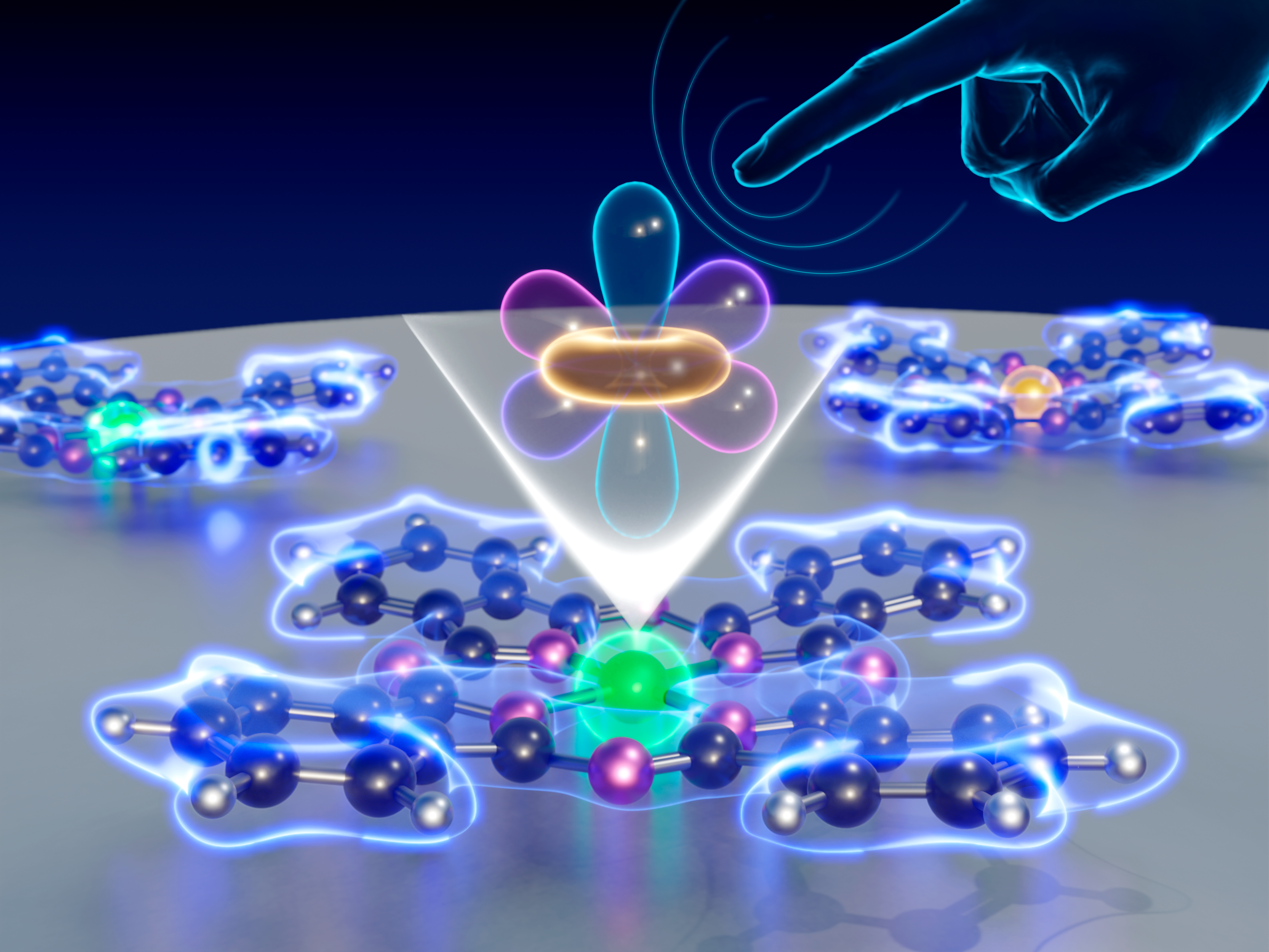
Observation of electron orbital signatures of single atoms within metal-phthalocyanines using atomic force microscopy
This illustration draws inspiration from Nan Yao’s groundbreaking research at Princeton University. Depicting a symbolic hand as atomic force microscopy (AFM), used to observe the electron orbital signatures of single atoms. This innovation has promising insights into electronic structures and mechanisms of chemical reactions.
Cryo-Electron Microscopy unveils Bacteriophage T7’s interaction with Escherichia coli
This artwork portrays the intricate interaction between bacteriophage T7 and Escherichia coli, focusing on the genomic DNA traversing the bacterial cell envelope. Based on cryo-electron microscopy data, the illustration shows the essential structural components of this process. Crafted in collaboration with Scixel (www.scixel.com), the artwork shows the groundbreaking research led by Jose L. Carrascosa’s group at CNB, shedding light on the dynamics of viral-bacterial interactions.
Interaction between capillary printed eutectic gallium alloys and gold electrodes
The cover art was inspired by the work of Navid Hussain, Michael Hirtz, and their team. It delves into the world of gold/liquid metal contacts, which are vital components in flexible electronics and other advanced technologies. With a keen focus on understanding the behavior of these contacts, the researchers employ a combination of cutting-edge chemical and morphological characterization techniques. The results shows new details on how these contacts evolve over time. Such insights are crucial for enhancing the performance and reliability of flexible electronics and hold the potential to drive innovations in a wide range of applications
Interfacial exchange phenomena driven by ferromagnetic domains
This cover illustrates the work of José Manuel Díez, Julio Camarero and coworkers in the field of magnetism. It centers around a phenomenon known as “exchange bias” in antiferromagnetic/ferromagnetic (AFM/FM) heterostructures. They found a new general mechanism that controls exchange bias, its temperature dependence. Surprisingly, it all comes down to the magnetic texture of the ferromagnetic layer during reversal. This newfound understanding not only allows for enhancing existing devices that rely on exchange bias but also opens up possibilities for designing innovative magnetic devices.
Innovative Cu3NbX4 nanocrystals enhance solar and photocatalytic performance.
The cover artwork is inspired by the research of Daniela R. Radu (Florida International University) and her collages. The artwork portrays the transformation of a 2D-TMDC nanosheet into niobium sulvanite nanocubes, denoted as Cu3NbX4 (where X = S or Se). These nanocubes exhibit immense potential as candidates for solar photovoltaics and photocatalytic water splitting, promising a sustainable energy future.
Tuning electronic and magnetic properties of lanthanide 2D networks via metal exchange.
This artwork illustrates a remarkable study on lanthanide multinuclear networks performed by Sofia O. Parreiras, David Écija, and their team (IMDEA Nanoscience). They showcase the ability to finely adjust the electronic and magnetic properties of these networks by exchanging the metal centers while keeping the same structural framework intact. By swapping the metallic centers from Erbium (Er) to Dysprosium (Dy), they observed a shift in the energy level alignment, which allowed them to tailor both the intensity and orientation of magnetic anisotropy. This breakthrough offers exciting prospects for designing advanced materials with tunable magnetic characteristics, presenting promising opportunities in the field of materials science and beyond.
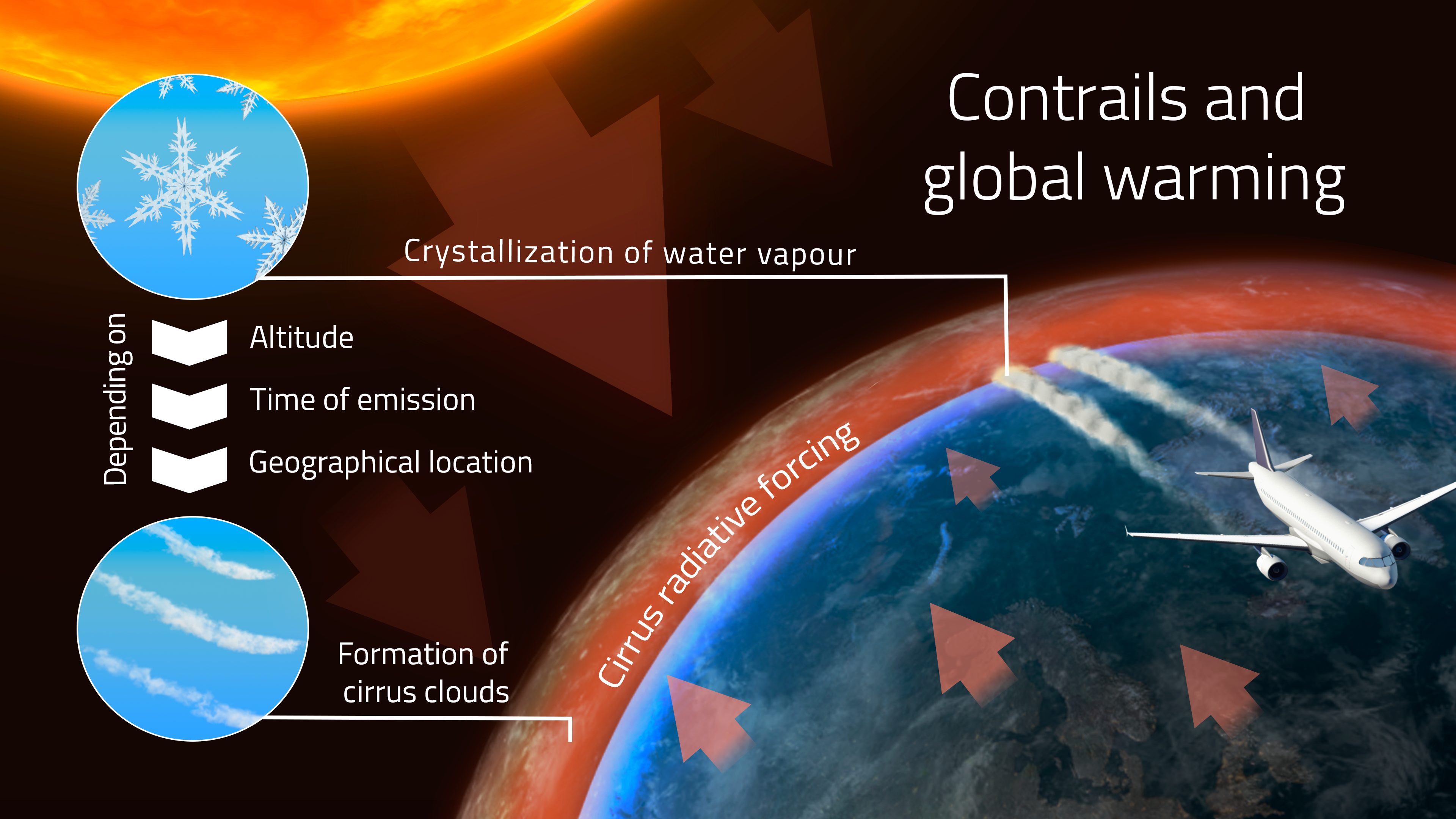
Contrails and Global Warming
This infographic, crafted for Manuel Soler at the PhD Program in Aerospace Engineering at the Universidad Carlos III, delves into the intricate relationship between contrails and global warming. Through visual storytelling, it explores the impact of contrails on Earth’s climate, offering insights into the complex interplay of atmospheric phenomena.
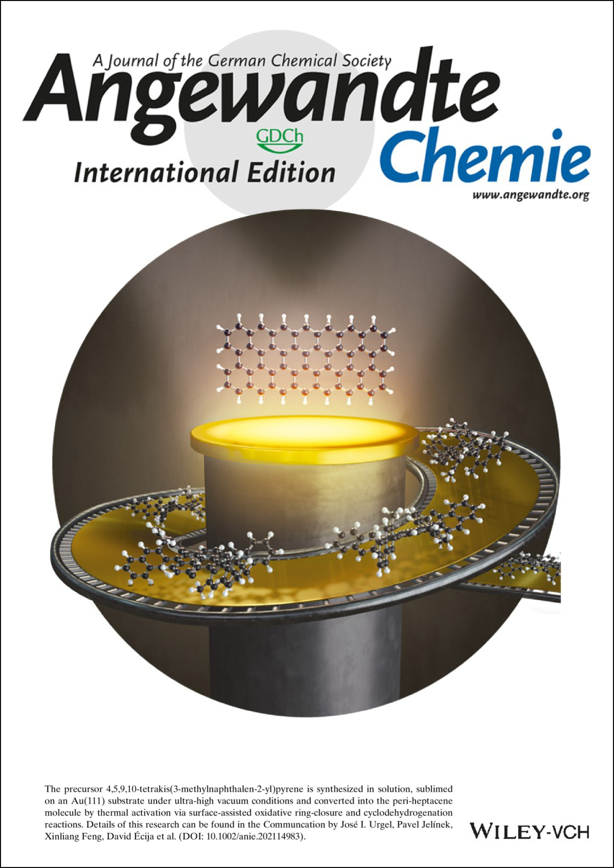
Synthesis and Characterization of peri-Heptacene on a Metallic Surface
This cover in Angewandte represents the research of , José I. Urgel, David Écija, (IMDEA Nanoscience) and their co-workers on synthesizing the precursor 4,5,9,10-tetrakis(3-methylnaphthalen-2-yl)pyrene in a solution. Subsequently, they sublimed it on an Au(111) substrate under ultrahigh vacuum conditions and converted it into the peri-heptacene molecule through thermal activation, utilizing surface-assisted oxidative ring-closure and cyclodehydrogenation reactions. This work sheds light on innovative approaches to creating complex molecules on surfaces, offering exciting prospects in the field of surface chemistry and materials science.
3D Imaging and Quantitative Subsurface Dielectric Constant Measurement Using Peak Force Kelvin Probe Force Microscopy
The illustration is based on the exciting work of Khaled Kaja and co-workers. Their study presents a novel method for measuring the dielectric constants of buried structures and interfaces. This technique combines peak force tapping quantitative Nano-mechanical mapping (PF QNM) with frequency-modulated Kelvin probe force microscopy (FM-KPFM). This innovative approach opens up new possibilities for 3D imaging, enabling detailed exploration of the dielectric constants in composite materials and heterostructures.
My thesis
This illustration holds a special place in my heart as it graces the cover of my thesis, a project that not only deepened my scientific understanding but also ignited my passion for visual communication. Through my research on protein aggregation and neurodegenerative diseases, I realized the profound impact of visual storytelling in science. It was a revelation—seeing complex concepts unfold visually, making them accessible and engaging, transformed my path, turning my hidden talent into a lifelong passion and my current profession.
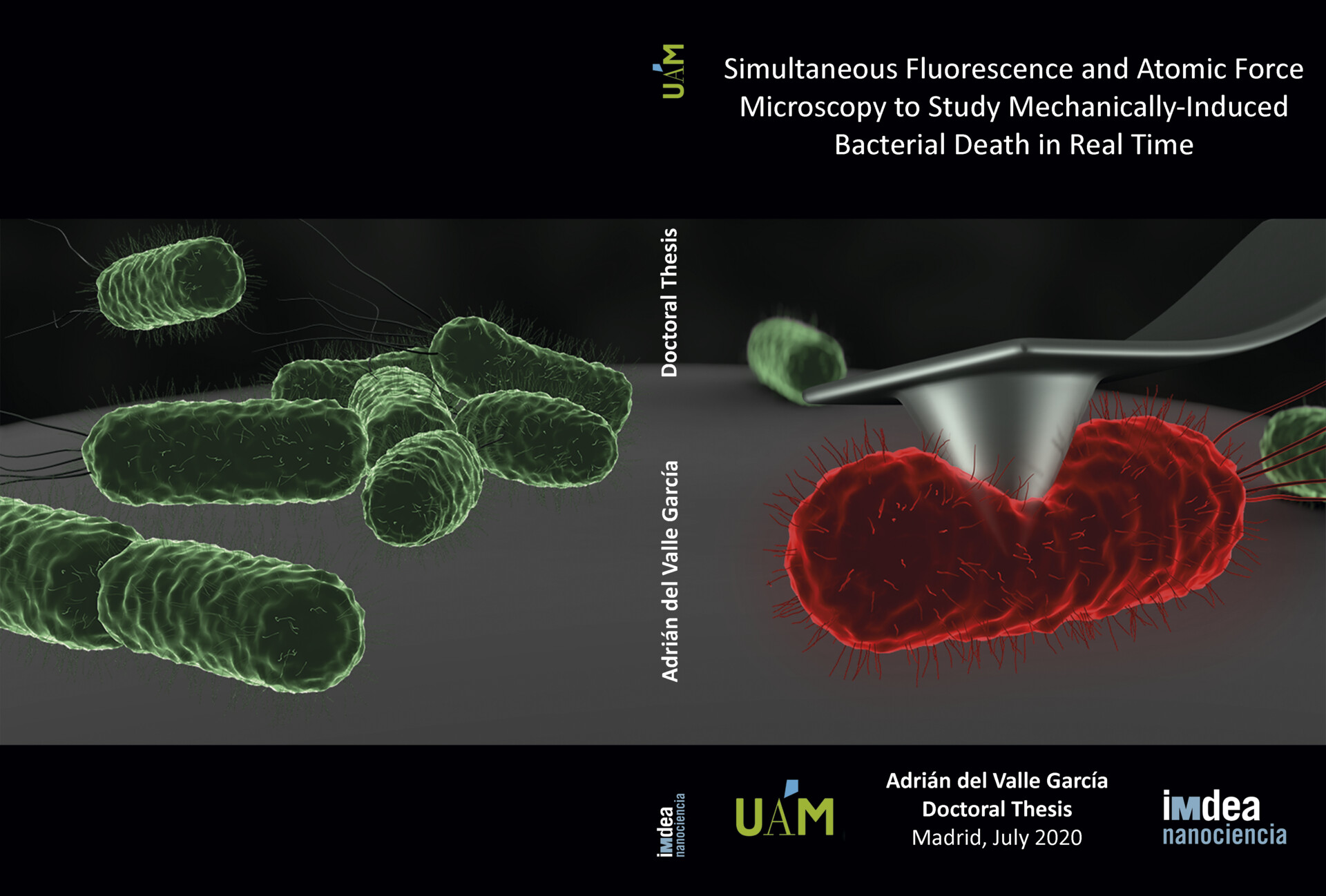
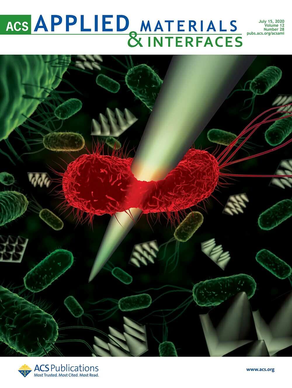
Simultaneous Fluorescence and Atomic Force Microscopy to study mechanically-induced bacterial death in real time
The purpose of this research was to kill bacteria using a nano-needle (cantilever tip of the Atomic Force Microscope) which records mechanical information useful for study parameters such as bacteria resistance, rigidity… And the death of the bacterium was imaged through fluorescence (using a reporter of membrane damage or of bacterial metabolism). This study is important for the design of new antimicrobial surfaces which are of great society interest since bacteria are more and more resistant to antibiotics becoming a world health problem.
Congratulation Adrian!
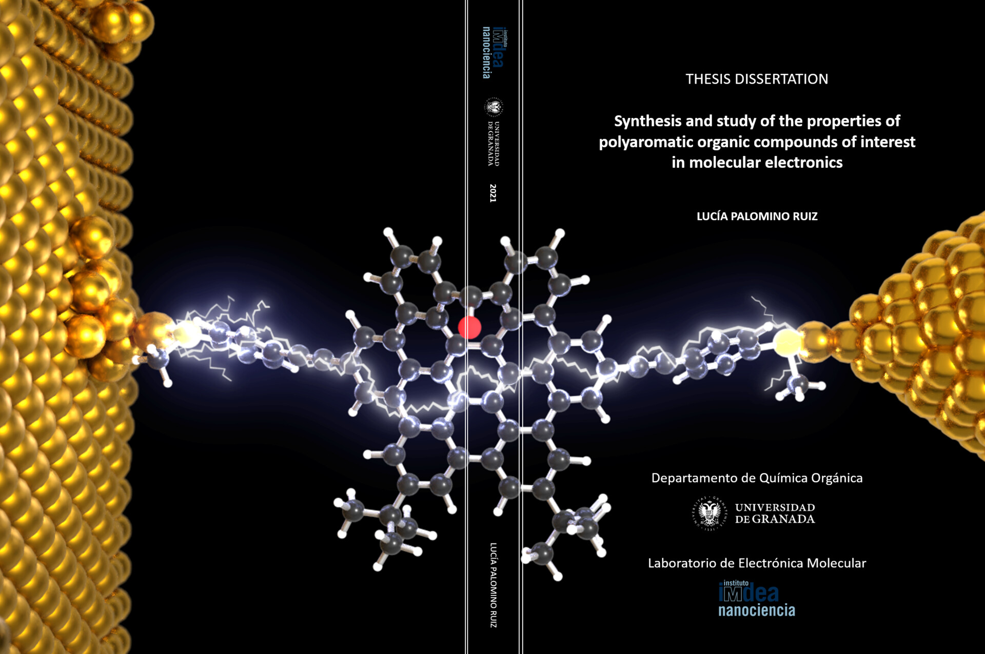
Synthesis and study of the properties of polyaromatic organic compounds of interest in molecular electronics
The illustration is an artistic representation of a molecular junction, created through the break-junction technique, where distorted hexabenzocoronene (HBC) derivative is wired between two gold electrodes.
Congratulations Lucía!
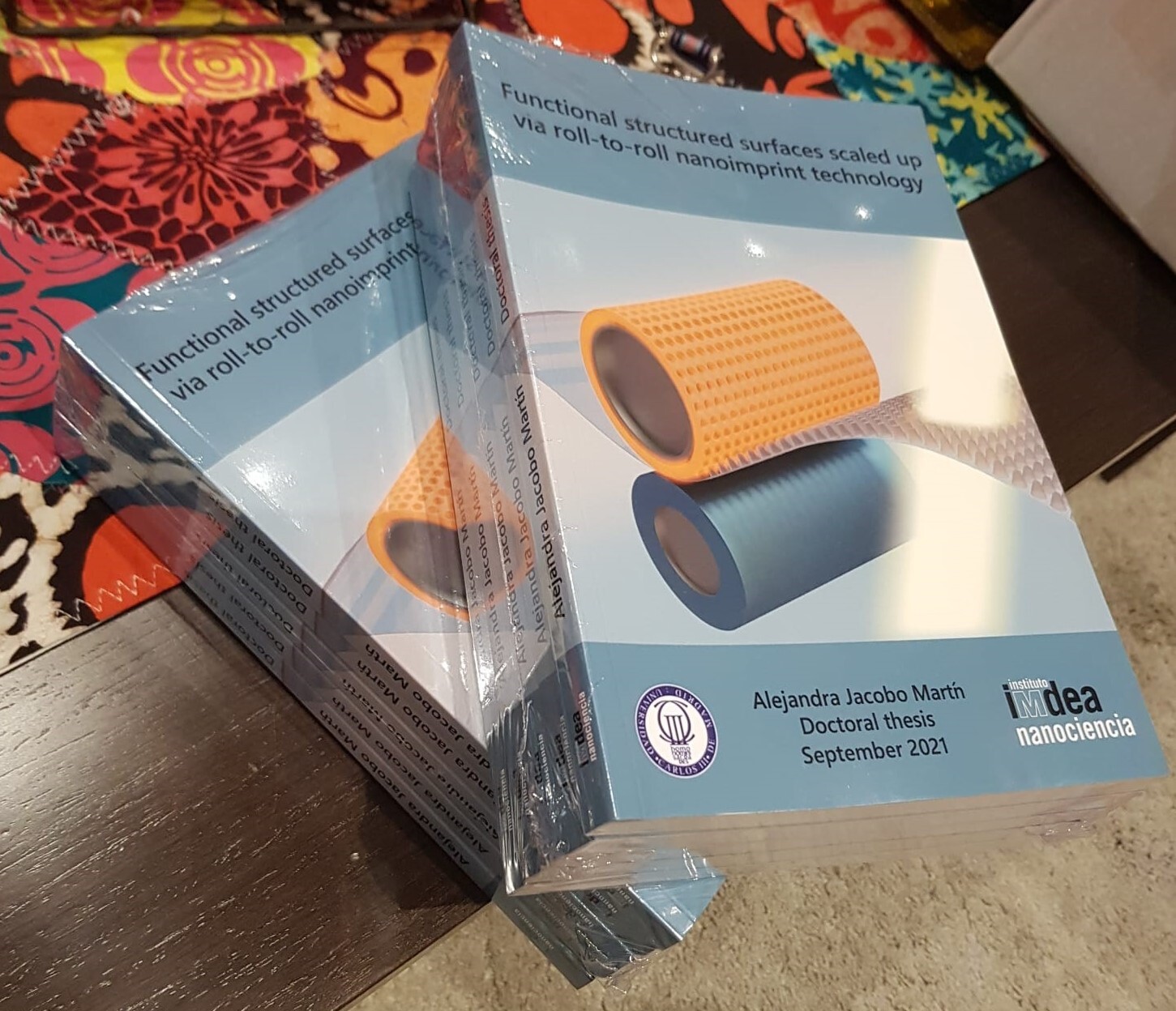
Functional structured surfaces scaled up via roll-to-roll nanoimprint technology
This image represents the large-scale production of nanoscale structures using roll-to-roll technology. This work is a step forward to produce low-cost antireflective flexible films optimized to reduce light losses.
Congratulations Alejandra!
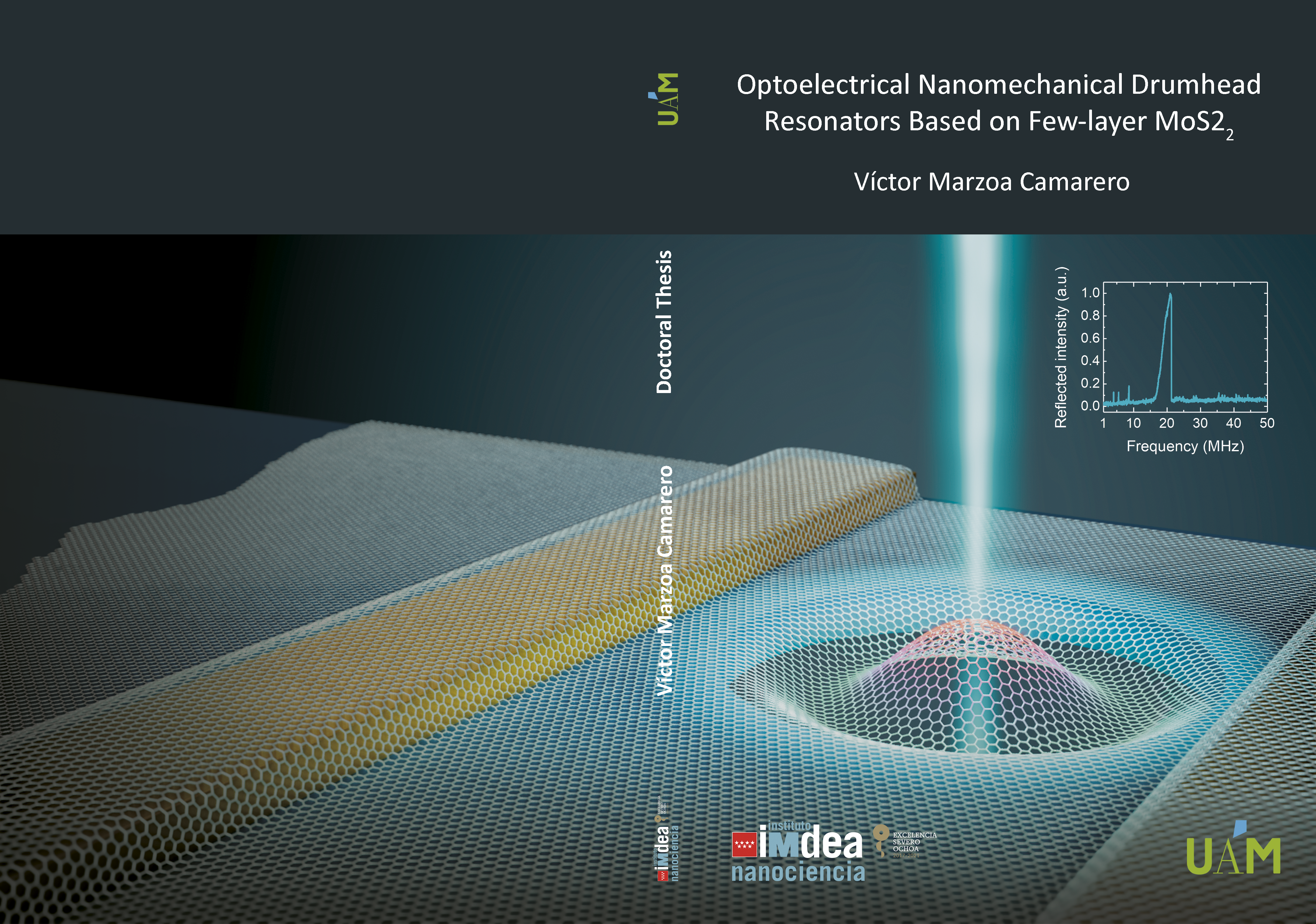
Optoelectrical Nanomechanical Drum Resonators Based on Few-Layer MoS2.
The image illustrates the work of Victor Marzoa in the development of optoelectrical nanomechanical drum resonators using few-layer molybdenum disulfide (MoS2). Through his work, he focuses on fabricating drum resonators incorporating MoS2 layers to investigate their unique optomechanical behavior.
Congratulations Victor!
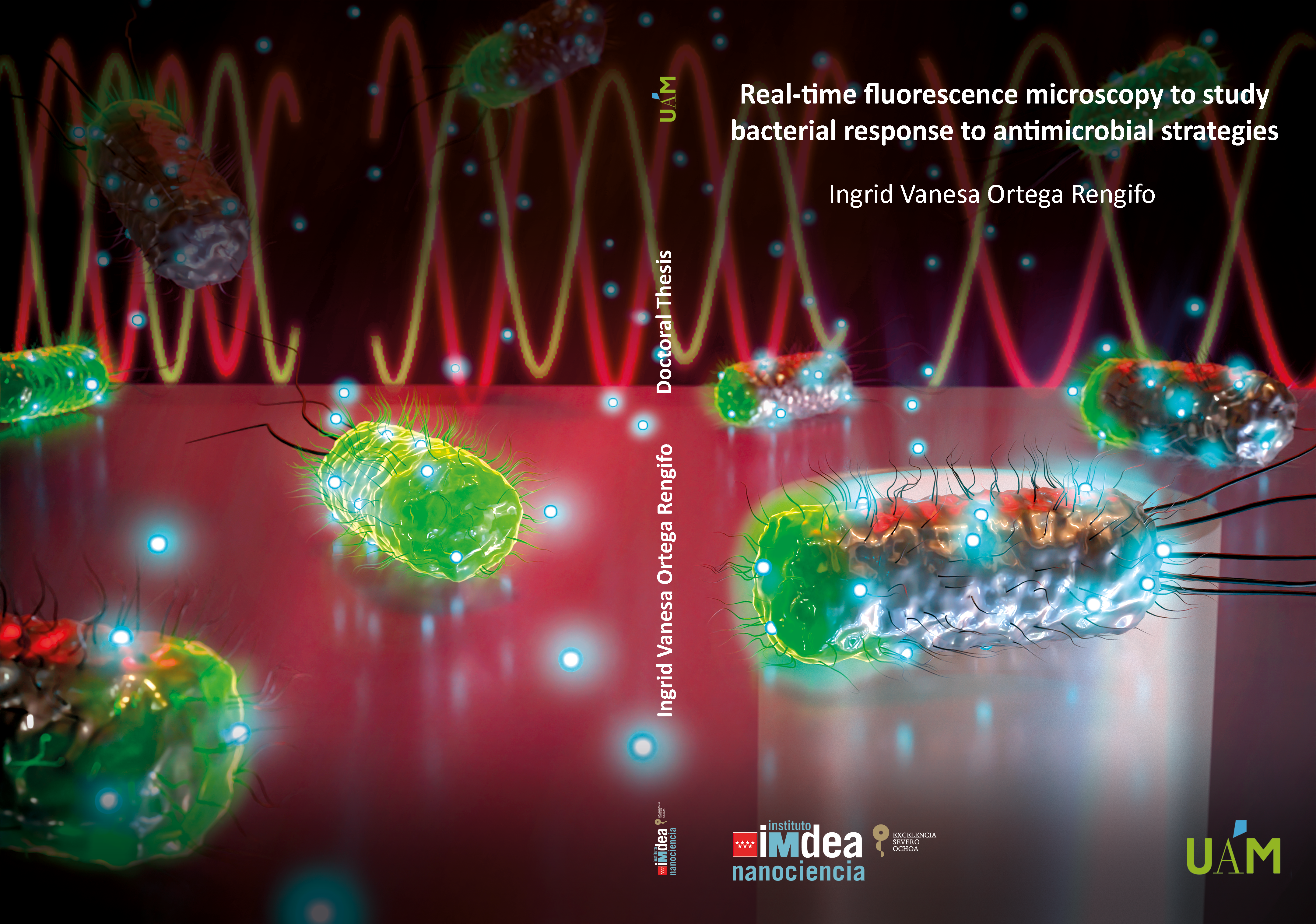
Real-time fluorescence microscopy to study bacterial response in antimicrobial strategies.
The cover art provides a visual representation of E. coli bacteria through the Min oscillations reporter. Using real-time microscopy, Ingrid Ortega is able to monitor the bacterial response to antimicrobial agents at the individual bacteria level. A key indicator of this response is the cessation of Min oscillations when the cell dies. Ortega’s research focuses on studying the effects of combining antimicrobials with photodynamic therapy. This artwork encapsulates the intricate dance of life and death at the microbial level, offering a glimpse into the innovative strategies being explored in the fight against bacterial infections.
Congratulations Ingrid!
Revolutionizing battery technology with fast-charging niobium oxide films
This artwork was created to illustrate the new research of Andrew Rappe, Arvin Arvin Kakekhani, Hyeon Han and coauthors. It was a collaborative effort from the University of Pennsylvania, Max Planck Institute, and University of Cambridge.
Since the 1940s, scientists have been studying niobium oxide, specifically T-Nb2O5, for creating efficient batteries. This material enables fast movement of lithium ions, leading to quicker charging. The challenge was growing high-quality, thin layers of T-Nb2O5. The authors achieved the growth of single-crystal T-Nb2O5 layers, facilitating rapid lithium ion movement. The material’s transitions in structure with changing lithium ion concentration allowed it to switch from an insulator to a metal, enhancing conductivity by a factor of 100 billion. By manipulating these transitions, they could control electronic properties, offering potential applications in high-speed battery charging and energy-efficient computing, among others.
Fabrication of molecular velcro: covalently bound MoS2–graphene heterostructures
Researchers at IMDEA Nanociencia, led by Enrique Burzurí and Emilio M. Pérez, achieved a pioneering breakthrough by covalently connecting 2D materials for the first time. The illustration shows this innovative approach, in which MoS2 and graphene layers are stitched using a bifunctional molecule. The resulting heterostructure’s electronic properties were dominated by the molecular interface. By combining MoS2’s semiconductor characteristics with graphene’s high mobility, the team constructed field-effect transistors that showcased modified gate voltage characteristics and suppressed current in graphene, attributed to the formation of covalent bonds. This achievement holds significant promise for tailoring electronic properties in 2D materials and advancing functional applications.
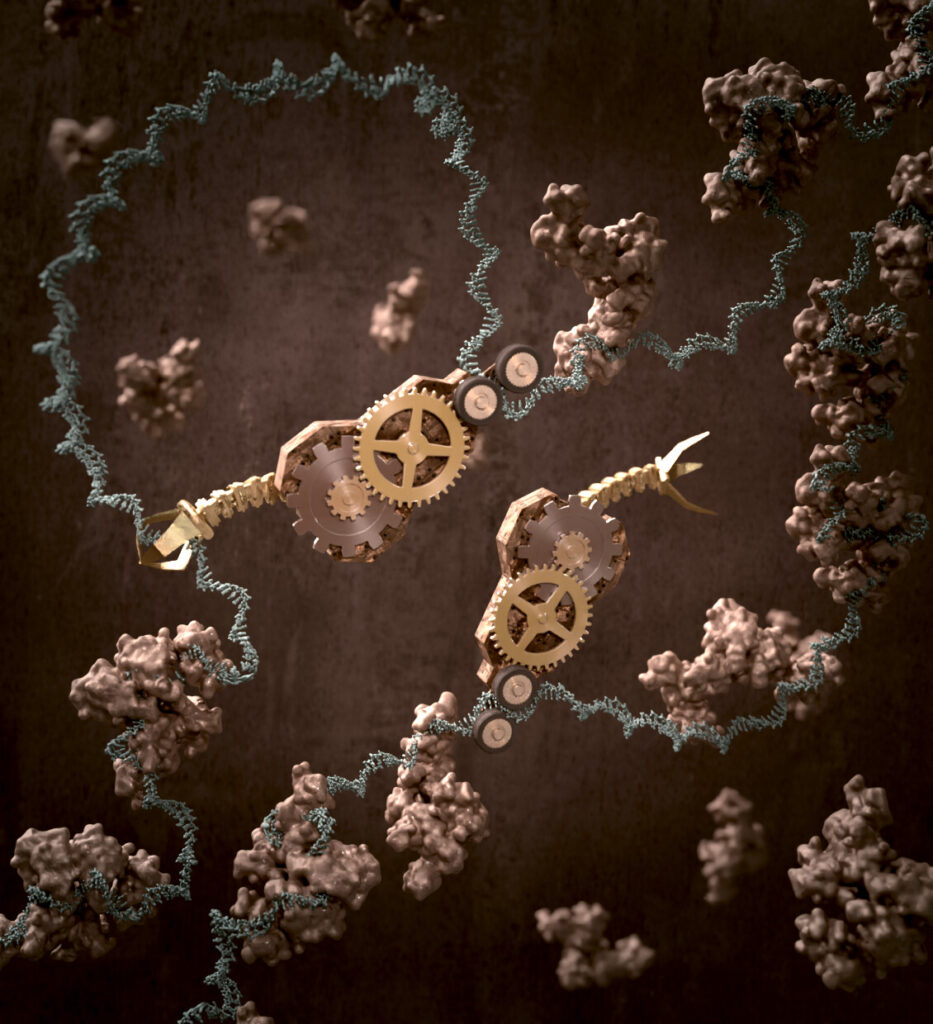
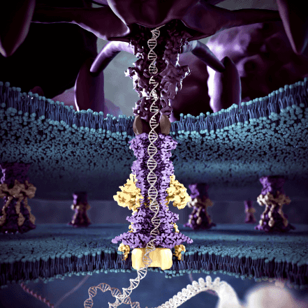
DNA helicase at work
This illustration is inspired by the research conducted by Fernando Moreno at the CNB and was a collaborative effort with Scixel (www.Scixel.com). The illustration depicts the HelB helicase enzyme in action on single-stranded DNA (ssDNA), showcasing a crucial process in DNA replication and repair. As HelB helicase unwinds the DNA double helix structure, it creates a loop in the ssDNA. This loop formation leads to the unbinding of other proteins, including single-stranded DNA binding proteins (SSBs), which play a pivotal role in stabilizing and protecting the single-stranded DNA regions during various DNA metabolic processes. The illustration captures the dynamic interaction between HelB helicase, ssDNA, and SSBs, highlighting the intricate molecular mechanisms at play during DNA unwinding and processing.

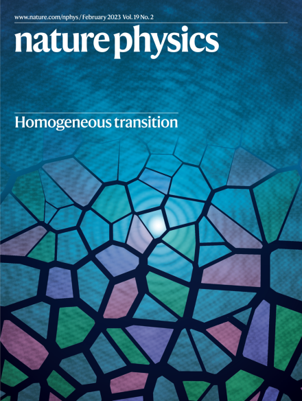
Neural networks for phase transition studies
This illustration visualizes the pioneering work of Julian Arnold and Frank Schäfer, led by Dr. Christoph Bruder at the University of Basel. It showcases a surface with magnetic material represented by the arrows and lens-like zoom that reveals a network of interconnected spheres and lines. This scene symbolizes their achievement in understanding phase transitions using neural networks, providing insights into complex systems. Their innovative approach to calculating optimal solutions without extensive training sheds light on the hidden workings of neural networks and their potential to detect phase transitions.

First direct observation of electron motion in action
This artwork, a collaborative effort with Scixel (www.scixel.com), was crafted to visualize the pioneering research conducted by Dr. Manish Garg’s group at the Max Planck Institute alongside scientists from the Autonomous University of Madrid and IMDEA Nanoscience. Researchers have achieved the remarkable feat of directly observing and comprehending electron movements within molecules on an attosecond scale. By bridging experimental techniques with theoretical insights, they managed to unveil the dynamic realm of electronic density in molecules.
New weapon against antibiotic-resistant bacteria: Light – activatable molecules
This scientific illustration captures the process of eradicating bacterial biofilms through photodynamic therapy. The concept highlights the use of a light-activated molecule that becomes toxic and emits fluorescence upon exposure to light, effectively targeting and destroying bacterial biofilms. This innovative approach underscores the potential of photodynamic therapy in combating microbial infections.
This artwork reflects my artistic interpretation of the research I conducted during my postdoctoral work with Cristina Flor’s group at IMDEA Nanoscience

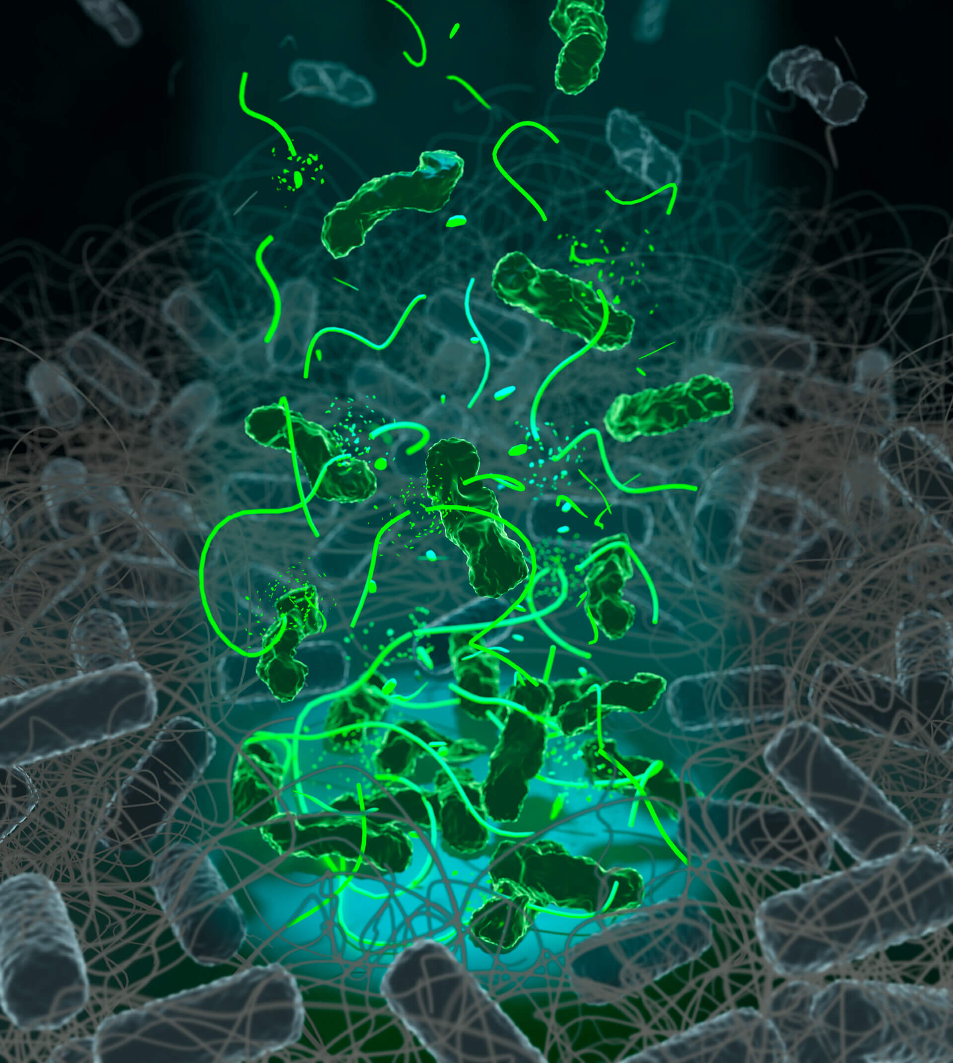
Biofilm, E.coli home sweet home
This illustration is my artistic representation of an Escherichia coli biofilm—a cave-like structure brimming with these bacteria. This complex formation serves as a protective fortress, shielding bacteria from antibiotics and the immune system, which makes infections particularly challenging to treat.
🖌️ Inspired by my postdoctoral work at IMDEA Nanoscience in Cristina Flor’s group, this artwork merges science and art to explore the hidden world of biofilms.

💡 Visualizing light-induced phase transitions in quantum materials 🔬
A new study of Allan S. Johnson and Daniel Pérez-Salinas, led by Prof. Simon Wall, reveals a groundbreaking imaging method that captures light-induced phase transitions in vanadium oxide (VO₂) at unprecedented spatial and temporal resolution. This innovation opens doors to engineering novel properties like superconductivity in quantum materials.
The illustration depicts a crystalline lattice melting (represented as a snowflake), superimposed with its coherent X-ray scattering pattern. It shows light inducing a change state from solid to liquid.
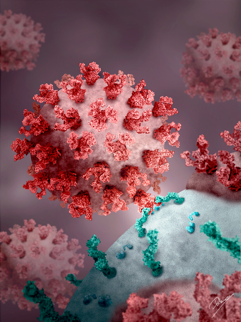
This illustration is a personal project, it represents the Human Immunodeficiency Virus (HIV) infecting a T-helper lymphocyte. The image shows the complex gp120 + gp41 of the HIV interacting with receptor CD4 and coreceptor CCR5 of the lymphocyte, one of the first steps of the viral infection.
In the host cell surface, we can observe fused rests of other viruses which have already entered the cell.
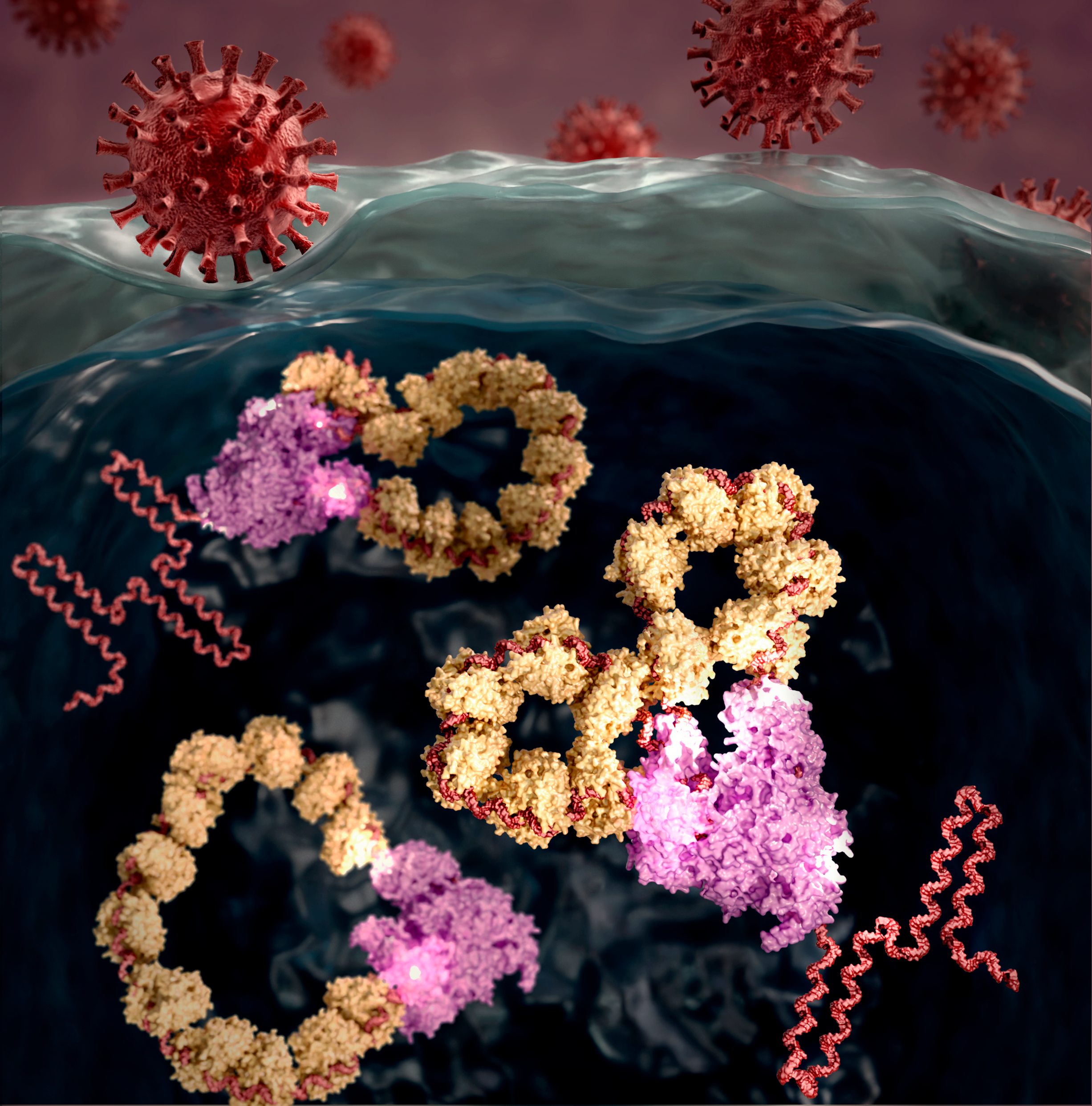
🔬 Visualizing influenza A virus replication at the nanometric scale 🦠
A groundbreaking study led by Borja Ibarra (IMDEA Nanoscience) and Jaime Martín Benito (CNB) has visualized how the influenza A virus multiplies its genome using high-speed atomic force microscopy. These findings reveal the intricate shape and movements of the viral macromolecules responsible for genome replication, enhancing our understanding of the virus’s multiplication cycle.
This illustration, created in collaboration with Scixel (www.scixel.com) captures the structure of the replicating virus system, showcasing the complexity of the viral machinery in action.
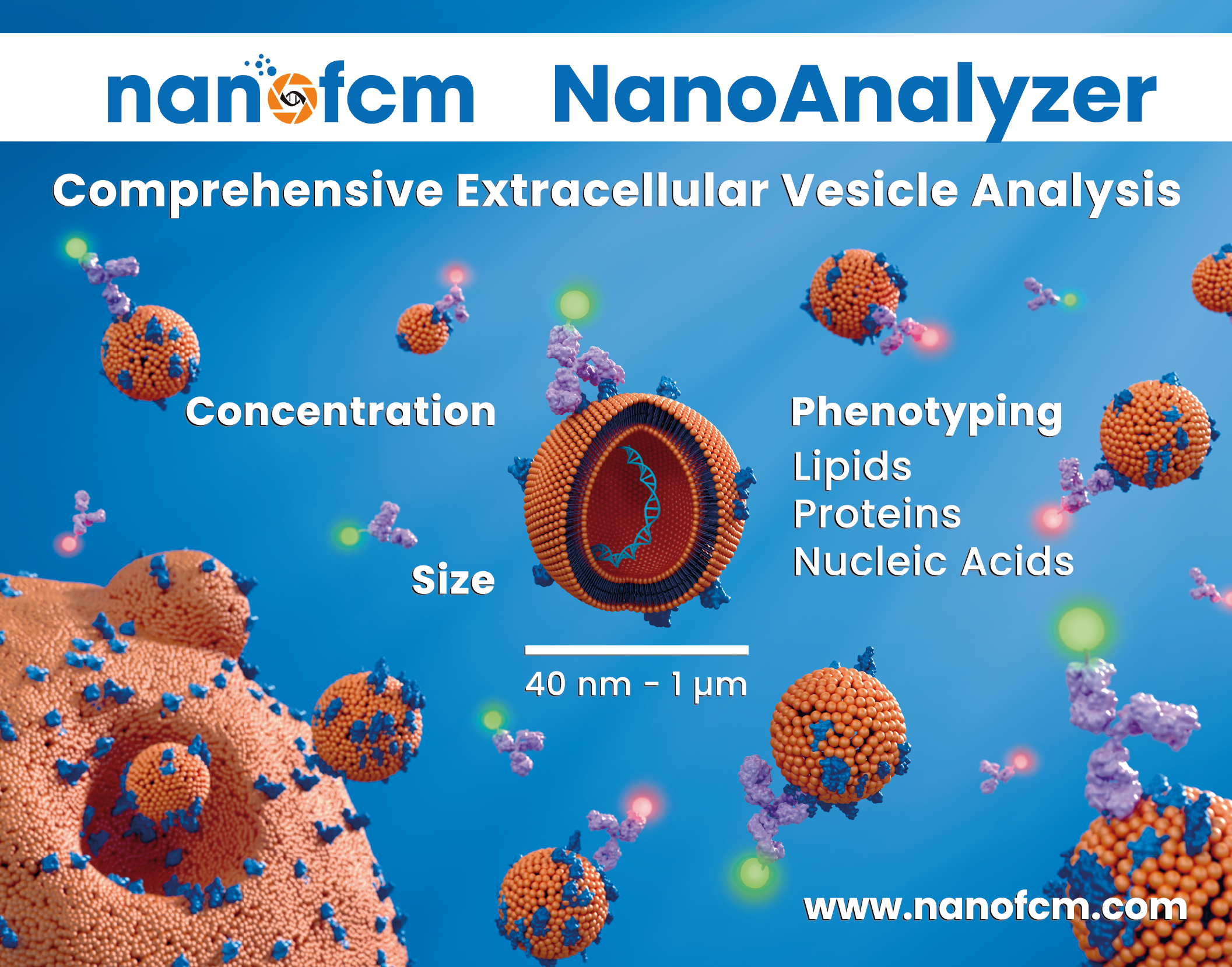
NanoFCM Banner
Artwork performed as a banner for NanoFCM (www.nanofcm.com) to promote their Nanoanalyzer as a powerful tool to characterize extracellular vesicles. The design was adapted for different international conferences.
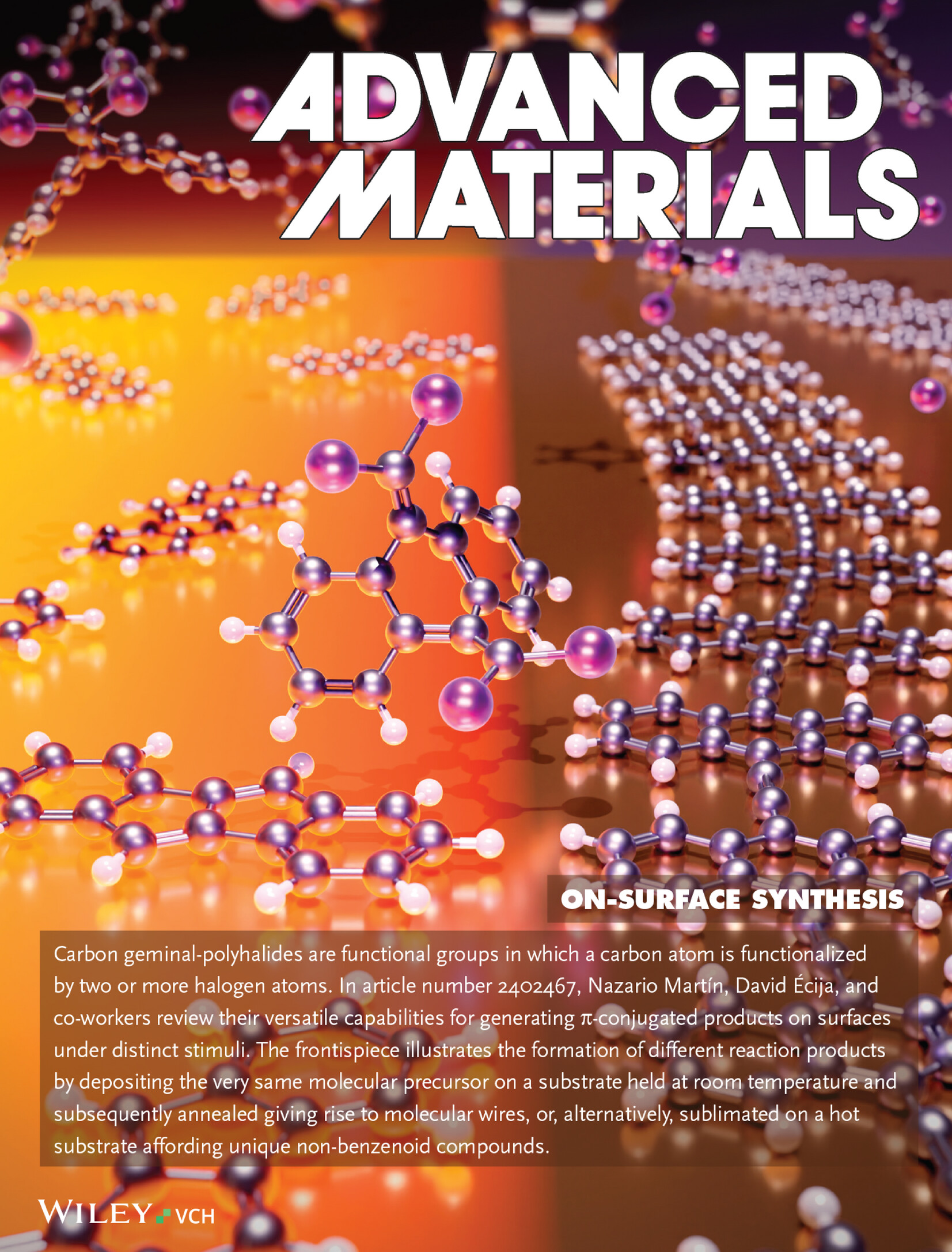
On-Surface Covalent Synthesis of Carbon Nanomaterials by Harnessing Carbon gem-Polyhalides
This image is inspired by the review by Nazario Marin, David Ecija, and their colleagues at IMDEA Nanoscience, showcasing the versatile capabilities of carbon geminal-polyhalides 🧬 in generating π-conjugated products on surfaces under distinct stimuli. It illustrates how the same molecular precursor can form different reaction products: deposition on a room-temperature🌡️ substrate is followed by annealing and creates molecular wires, while direct sublimation onto a hot substrate 🔥 yields unique non-benzenoid compounds.
Turning sunlight into hydrogen energy
Wahid Ullah, mohamed nawfal Ghazzal and his team have downsized the graphdiyne (GDY) network to shape quantum dots (QDs), unlocking its optical properties! ✨These tiny GDYO-QDs absorb light across a wide range, making them perfect for converting sunlight into hydrogen fuel, a clean energy source 🌍. By combining GDYO-QDs with TiO2, the team tremendously boosts the photocatalytic performance, opening new doors for using GDYO-QDs in future energy technologies.
⭐Empowering CO2 Eco-refrigeration⭐
Did you know that the traditional refrigeration processes contribute significantly to global warming?🌡️This illustration represents recent research where scientists have proven that MOF-508b is an excellent material for eco-friendly🌿 cooling and heating systems. Currently, 20% of our electricity goes to refrigeration and 50% to heating. This study shows how MOF-508b, a revolutionary material, uses CO2 as an eco-friendly refrigerant. At low CO2 pressures, it can heat up to 56°C and cool down to -10°C, making it ideal for space heating, air-conditioning, and food refrigeration. This cover was created with Agata Communications.
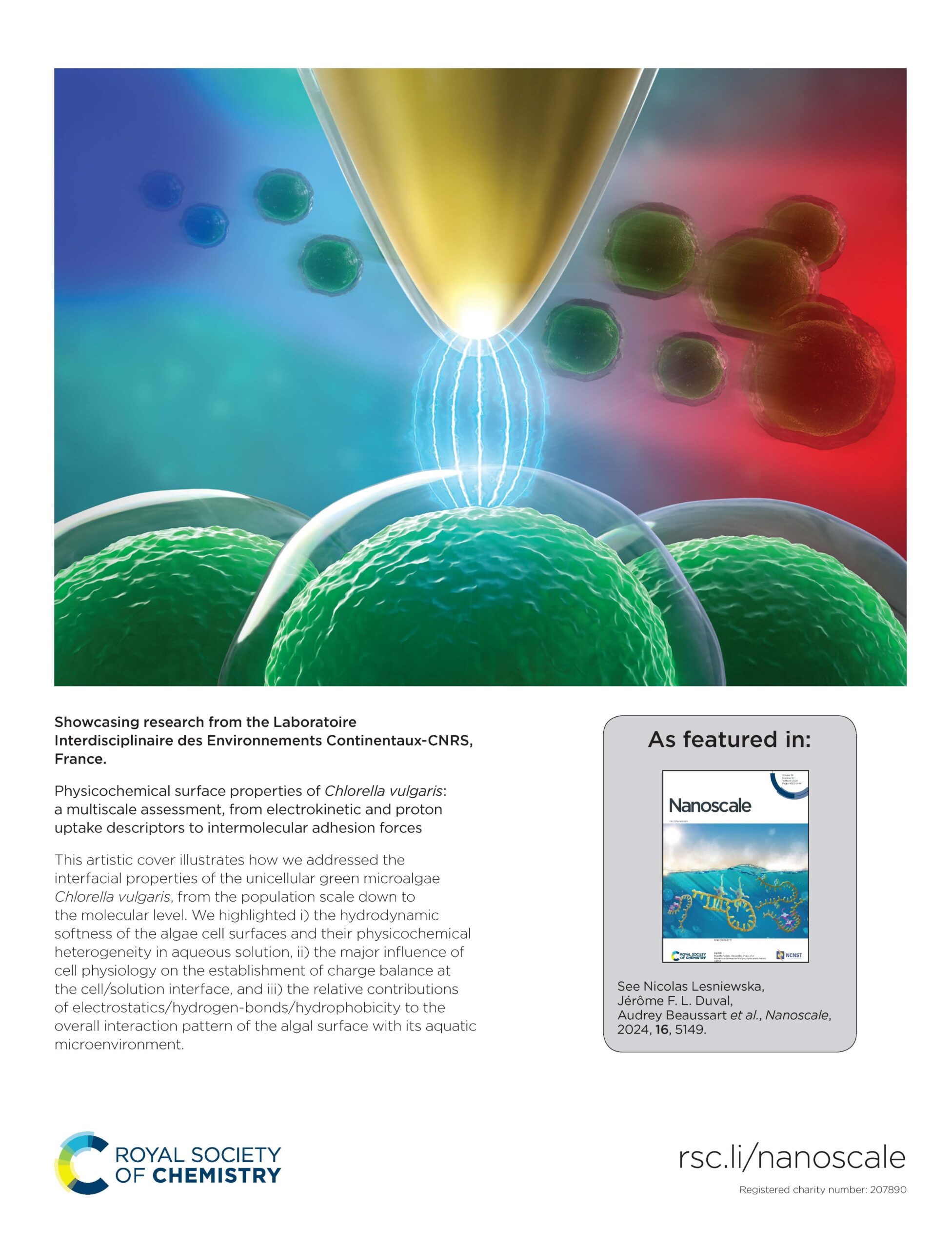
🔓Unlocking microalgae potential! 🌿✨
This image is inspired by the research of Nicolas Lesniewska, Audrey Beaussart and co-workers. They study the physicochemical surface properties of microalgae, a key to unleashing its potential in biotechnological industries and green energy. 🌱 By using electrophoretic analysis, the team has shed light on how microalgae interact within various environments, revealing the dynamic nature of their surfaces. In the illustration, the 🔵 and 🔴 spots represent the negative and positive electrodes, respectively, which create an electric field. This field provokes the migration of algae, providing insights into their surface charge characteristics.🔬👀 To get a close-up view, the authors used Atomic Force Microscopy (represented by the golden tip), unveiling the heterogeneity on the surface of individual algae cells.
Today’s science is the boat that sails us into tomorrow’s future
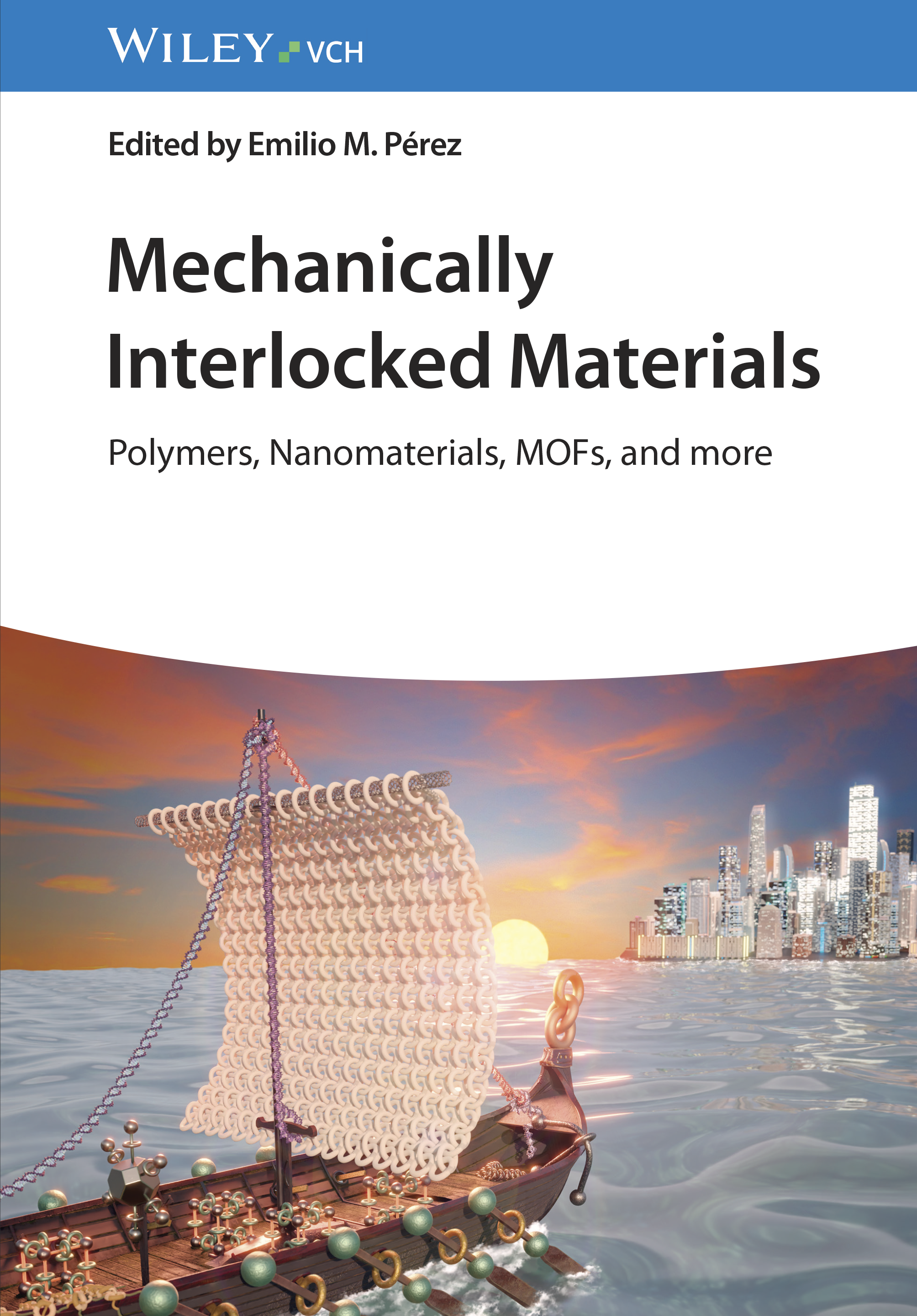
The image features a boat formed by mechanically interlocked molecules (MIMs) and materials (MIMats), sailing towards a futuristic skyline. This visual metaphor captures the essence of the book, illustrating how these advanced materials are navigating us towards new horizons in science and technology. This artwork was commissioned by Emilio M. Pérez to illustrate the book “Mechanically Interlocked Materials: Polymers, Nanomaterials, MOFs and more”
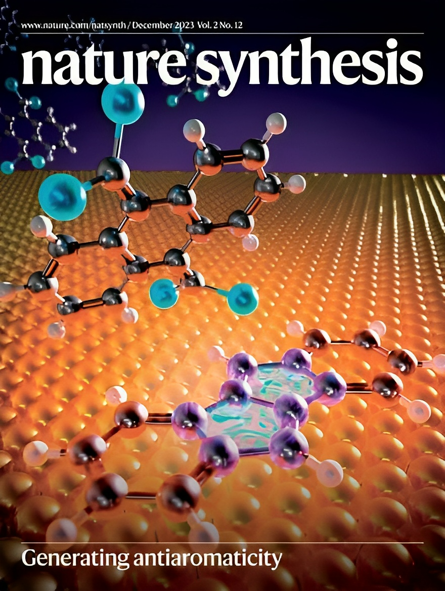
Innovative strategy for synthesizing antiaromatic compounds
Face to face, the precursor and its future self – an antiaromatic polycyclic conjugated hydrocarbon – gaze upon each other. A depiction of ‘what is’ and ‘what will be’ as soon as the precursor meets the hot metal surface.
An artwork commissioned by David Écija and his team to illustrate their new publication on Nature Synthesis.
Hybrid metrology revolutionizes the analysis of nanostructured metasurfaces.
The image illustrates a cylindrical array of TiO2 nanostructures used as a model system for developing a hybrid metrology strategy. It integrates synchrotron-based and lab-scale analytical techniques, providing new insights into the optical properties of metasurfaces. This work was inspired by the research of Irdi Murataj, Eleonora Cara, and their colleagues of the Istituto Nazionale Ricerca Metrologica (INRiM),
What’s similar between a butterfly and molecular orbital configuration?
This image was created to illustrate the work of Saül Garcia-Orrit , Matteo Tommasini, Juan Cabanillas and co-workers. Their research reveals the photophysics of a nanographene metalloporphyrin. It was very inspiring to work with Saül Garcia-Orrit, who came up with the beautiful concept of linking the movement of the butterfly wings of Rafael Araujo with the different open and close positions of the molecular orbital configurations.
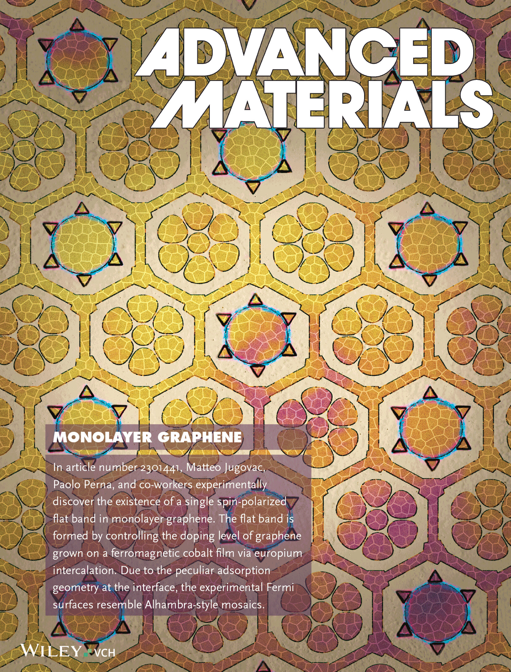
Alhambra-style mosaics
Matteo Jugovac, Paolo Perna, and their team discovered a single spin-polarized flat band in monolayer graphene, capable of unlocking exotic electronic and magnetic states like unconventional superconductivity and quantum phases. By doping graphene on cobalt with europium, they achieved spin-polarized flat bands that resemble Alhambra-style Fermi surfaces. This innovative approach combines spintronics with graphene’s unique properties, revealing new avenues for advanced spin-based electronic devices and highlighting europium’s crucial role in modifying graphene’s structure and enhancing interlayer magnetic coupling for topological materials.
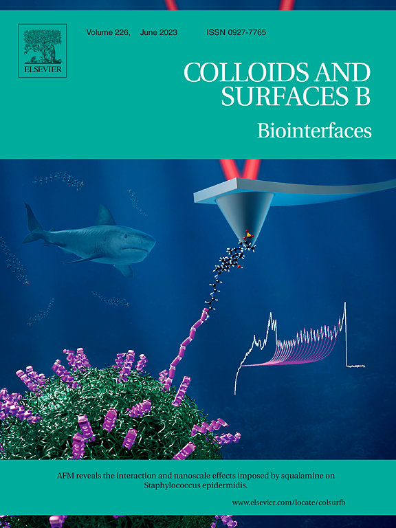
AFM reveals the interaction and nanoscale effects imposed by squalamine on Staphylococcus epidermidis
This coverart illustrates the research conducted by Sofiane EL-Kirat-Chatel, Audrey Beaussart and coworkers at CNRS. Their study focused on exploring the antimicrobial effects of squalamine, a natural compound derived from dogfish sharks, on the troublesome Gram-positive bacterium, Staphylococcus epidermidis. Using atomic force microscopy (AFM), the researchers revealed the binding mechanism of squalamine to the bacterial cell surface, driven by electrostatic interactions. This work demonstrated the utility of AFM and microbiological assays in unraveling the molecular mechanisms underlying squalamine’s antibacterial properties.
Experimental demonstration of a magnetically induced warping transition in a topological insulator mediated by rare-earth surface dopants
Beatriz Muñiz Cano, Miguel A. Valbuena, and their colleagues from IMDEA Nanoscience have spearheaded research into magnetic topological insulators, a special type of material with unique properties. These materials exhibit a fascinating interplay between surface states and magnetic order. This combination leads to a change in the symmetry of these materials, predicted to alter their shape from a hexagon to a triangle when a certain energy gap is opened.
The cover art illustrates this transition, which was observed in a specific type of these materials, called Bi2Se2Te, when doped with rare-earth elements like Erbium (Er) and Dysprosium (Dy). The authors also noticed that by increasing the amount of these rare-earth elements, the properties of the surface states can be controlled, allowing to gradually adjust the energy level towards the opened gap.
These findings open up new ways to control the interaction of magnetic fields with these surface states, and could potentially lead to the realization of a quantum phenomenon known as the quantum anomalous Hall effect.
Ultrafast X-ray imaging of the light-induced phase transition in VO2
What unfolds when we delve into the very early stages of phase transitions? This is the intriguing question that Simon Wall and his coauthors sought to answer using ultrafast techniques. The cover art provides a visual narrative of their research into the intermediate states of photo-excited phase transitions. Conventionally, these states are expected to be inhomogeneous, but their research paints a different picture. Employing a spatially resolved ultrafast X-ray imaging technique, they discovered that the initial stages of the metal-insulator transition in VO2 are, in fact, homogeneous. The anticipated inhomogeneity only emerges after a few hundred femtoseconds. This unexpected revelation challenges traditional paradigms and paves the way for novel explorations in the field of phase transitions.
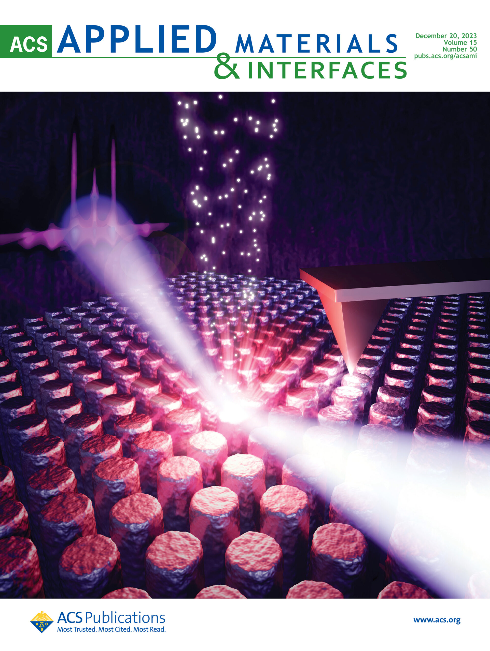
Characterizing sequential infiltration synthesis in polymers through X-ray analysis.
This cover art illustrates Dr. Eleanora Cara and her co-author’s research. It shows the ex-situ grazing incidence X-ray fluorescence quantification technique applied to polymers and block copolymers infiltrated with inorganic compounds during the sequential infiltration synthesis process. The illustration depicts how the X-ray beam, upon impinging on the materials, excites fluorescence in a specific target atom, such as aluminum atoms in alumina deposited using trimethyl aluminum and water as gas-phase precursors. This fluorescence can then be harnessed for the absolute quantification of the mass concentration of the compound.
Infrared-activated bactericide: rhenium disulfide (ReS2)-functionalized mesoporous silica nanoparticles
This cover artwork illustrates the research of Cheng-Yu Lai and coworkers (Florida International University) exploring the potential of multifunctional nanomaterials in the fight against antibiotic-resistant bacteria. The study highlights the synergetic effect of combining antibiotic chemotherapy and photothermal therapy (PTT) in combating bacterial infections. It shows the antimicrobial power of rhenium disulfide (ReS2)-functionalized mesoporous silica nanoparticles (MSNs) under near-infrared laser illumination. The MSN-ReS2 nanoparticles, with their enhanced photothermal efficiency and controlled drug delivery capability, demonstrate over 99% bactericidal efficiency against both Gram-negative and Gram-positive bacteria upon laser irradiation. Remarkably, when loaded with tetracycline hydrochloride, these nanoparticles achieve a 100% bactericidal effect against E. coli.
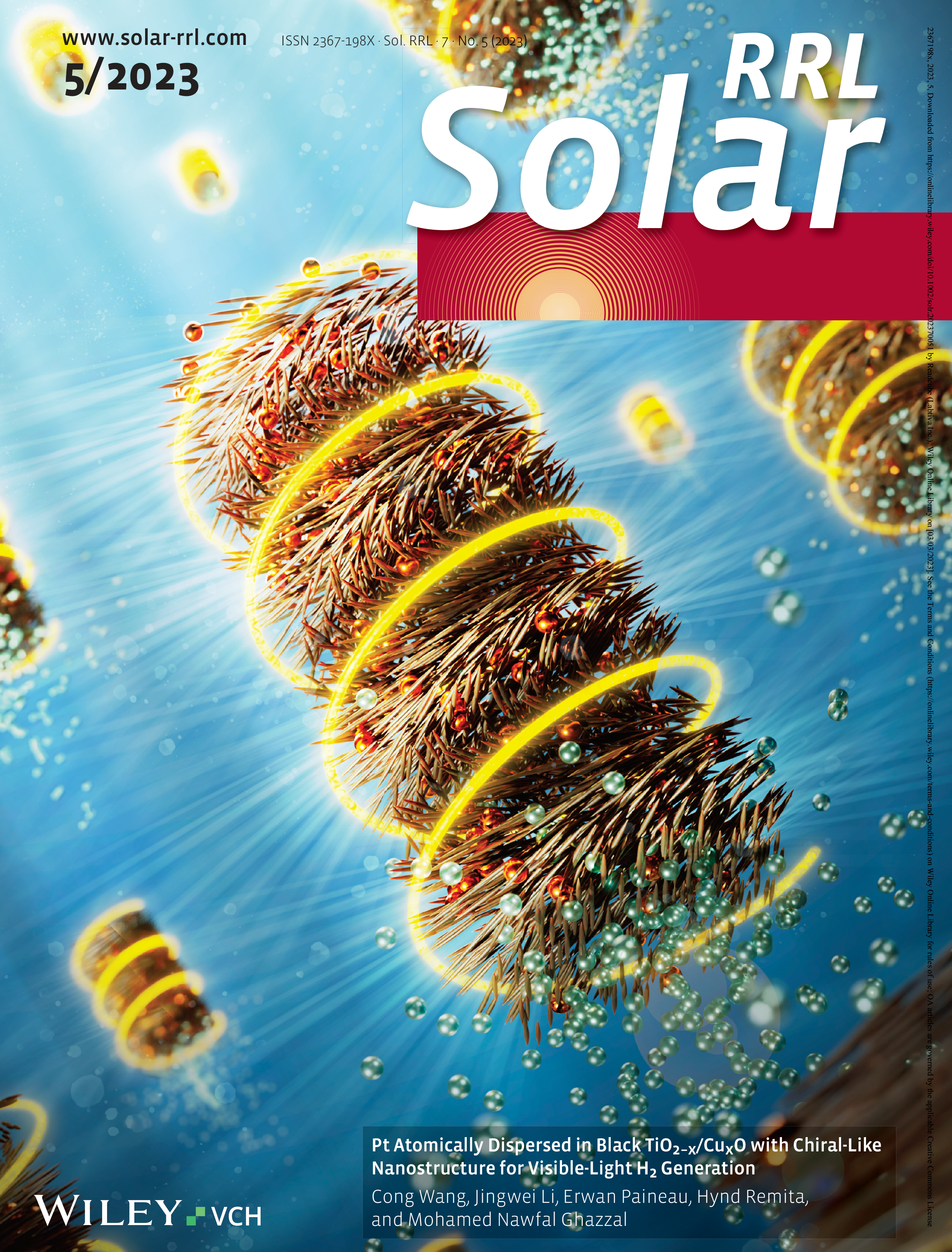
Chiral materials enhance light capture for better hydrogen production
This cover illustrated the research of Mohamed Nawfal Ghazzal and his team (Institut de Chimie Physique-CNRS) about hydrogen generation. They designed a material called black TiO2−x/CuxO with chiral-like structures, where a single Pt atom was successfully deposited on both black TiO2−x and CuxO to enable visible-light hydrogen production. This innovative work offers valuable insights into the rational construction of nanostructured materials featuring chiral nematic structures, which have the potential to greatly enhance light harvesting capabilities. The study opens new possibilities for advancing the field of renewable energy through efficient hydrogen generation using sunlight.
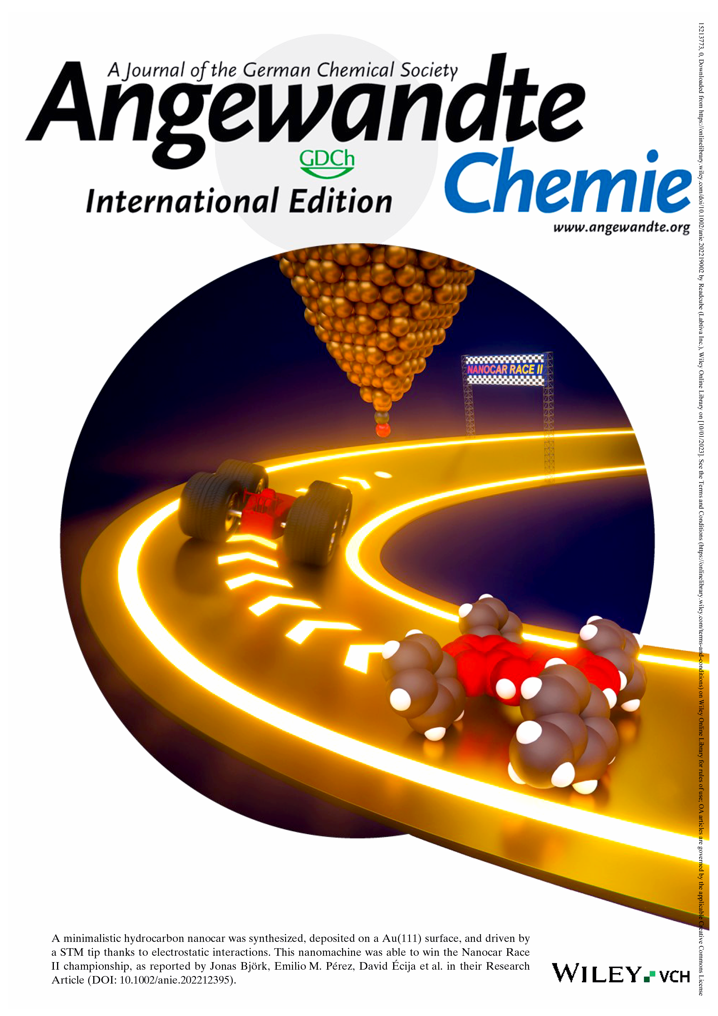
Design and manipulation of a nanocar
This artwork depicts the winning nanocar of the Nanocar Race II competition. Created as a cover for Angewandte Chemie, it celebrates the remarkable research of Emilio M. Pérez, D. Écija and co-workers. The nanocar of the illustration is inspired by the molecule of Nanohispa Team, whose design propelled them to victory in the Nanocar Race II. The paper details the molecular design and construction of this cutting-edge nanocar, featuring an anthracene chassis and four benzene derivative wheels. Sublimated and adsorbed on an Au(111) surface, the nanocar demonstrates controlled and agile manipulation using the tip of a scanning tunneling microscope (STM).
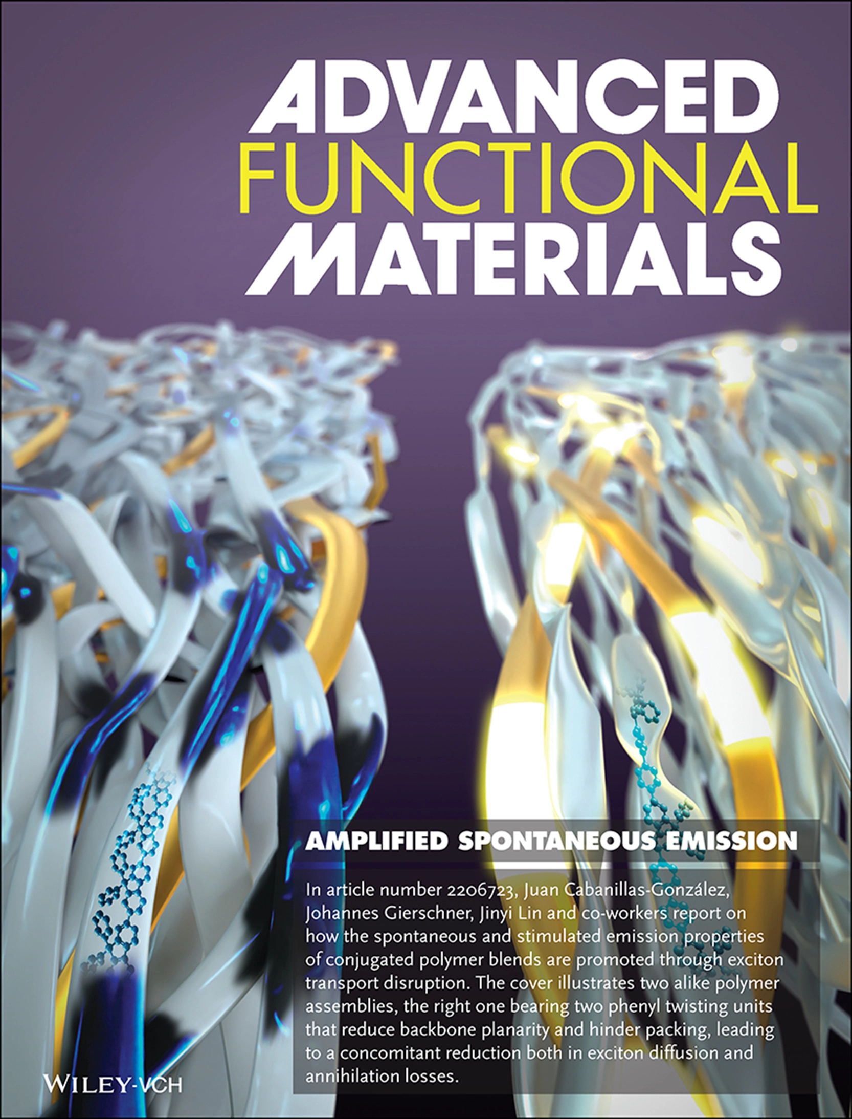
Twisting polymers to boost stimulated emission properties in lasers
Researchers led by Juan Cabanillas-González present an exciting study on how the properties of conjugated polymer blends can be enhanced. They found that by disrupting exciton transport, the spontaneous and stimulated emission of these polymers is significantly improved. The cover art showcases two polymer assemblies, one on the right with two phenyl twisting units. These units reduce the planarity of the polymer backbone and hinder the packing, leading to a decrease in both exciton diffusion and annihilation losses. This shows the different ways in which molecular arrangements can influence the behavior of polymers, offering exciting prospects for advancing optoelectronic technologies. The research provides valuable insights into the design and optimization of conjugated polymer materials, which will help to develop new applications in energy-efficient light-emitting devices and other cutting-edge technologies.

Exploring the anticancer potential of reactive osmium CN complexes
Sonia Infante, Ana Pizarro, and her team at IMDEA Nanoscience have achieved a significant breakthrough. By employing an innovative strategy, they developed highly potent anticancer Os-arenes by chelating anionic C,N-phenylpyridine ligands. This contrasts strikingly with previously reported analogues featuring N,N-bipyridine ligands. The outstanding intracellular reactivity of these Os-arenes can be attributed to two crucial factors: the facile aquation of the Os–Z bond and the elevated basicity of the Os-coordinated water molecule. This discovery, which served as inspiration for the cover artwork, sheds light on the unique mechanisms underlying the anticancer properties of these Os-arenes. These findings open opportunities for potential therapeutic innovations with enhanced efficacy.
Bacterial degrons in synthetic circuits
The research led by Javier Buceta, Arantxa Urchueguía, and their colleagues at South Dakota State University delves into the promising realm of bacterial proteases in synthetic circuits, which inspired this cover art. Bacterial proteases (Pac-Man), which recognize specific amino acid degradation tags known as “degrons” (in blue), offer a promising strategy for post-translational regulation. These degrons can be fine-tuned to control the degradation levels of tagged proteins, making them a focal point in recent research endeavors. The team’s work provides a comprehensive review of the latest applications of degradation tags for circuit engineering in bacteria.
Bioinspired nanocone surfaces offer a new strategy to combat bacteria
Researchers from IMDEA Nanoscience, led by María Teresa Alameda and Isabel Rodríguez, have unveiled a powerful strategy for combating bacteria using a “fakir” surface. The cover artwork represents how bacteria undergo deformation upon attaching to bioinspired bactericidal nanocone surfaces. The researchers devised an innovative and straightforward method by flipping the bacteria over, enabling them to observe and measure the indentation caused by the attachment using cutting-edge techniques like atomic force microscopy and stereometric analysis of scanning electron microscopy images. Through this approach, valuable insights into the interaction between bacteria and the nanocone surfaces were gained, setting the stage for potential applications in the design of highly effective bactericidal materials. This research significantly advances our understanding of bacterial behavior and holds promise for creating novel antimicrobial strategies and improving bioinspired surfaces for diverse practical uses.
Interplay between π-conjugation and exchange magnetism in one-dimensional porphyrinoid polymers
The cover artwork showcases a new research breakthrough performed by José I. Urgel and colleagues from IMDEA Nanoscience. In this study, scientists successfully formed one-dimensional porphyrinoid polymers by connecting individual units through a unique process called [3 + 3] cycloaromatization using isopropyl substituents. What’s interesting is that these connected units showed intriguing magnetic behavior. Specifically, the spins of adjacent porphyrinoid units interacted in a way called “antiferromagnetic coupling,” where the closest spins had opposite magnetic effects. This finding highlights the importance of a special type of molecular arrangement, called π-conjugation, in influencing how the magnetic properties of these materials are interconnected. These discoveries provide valuable insights into the behavior of these molecular structures and their potential applications in future technologies.
How Much Force Is Needed To Kill a Single Bacterium?
The illustration was crafted for Cristina Flors’ research group at IMDEA Nanoscience. It was the centerpiece of the thesis of my dear colleague, Adrian del Valle. It portraits the Atomic Force Microscope (AFM) as a needle, deftly piercing through the bacterium’s membrane. This breakage of the bacterial membrane facilitates the entry of a fluorescence dye, which exhibits red fluorescence when it binds to DNA and is illuminated with green light. As a result, the optical microscope can detect damaged bacteria through this fluorescence response.
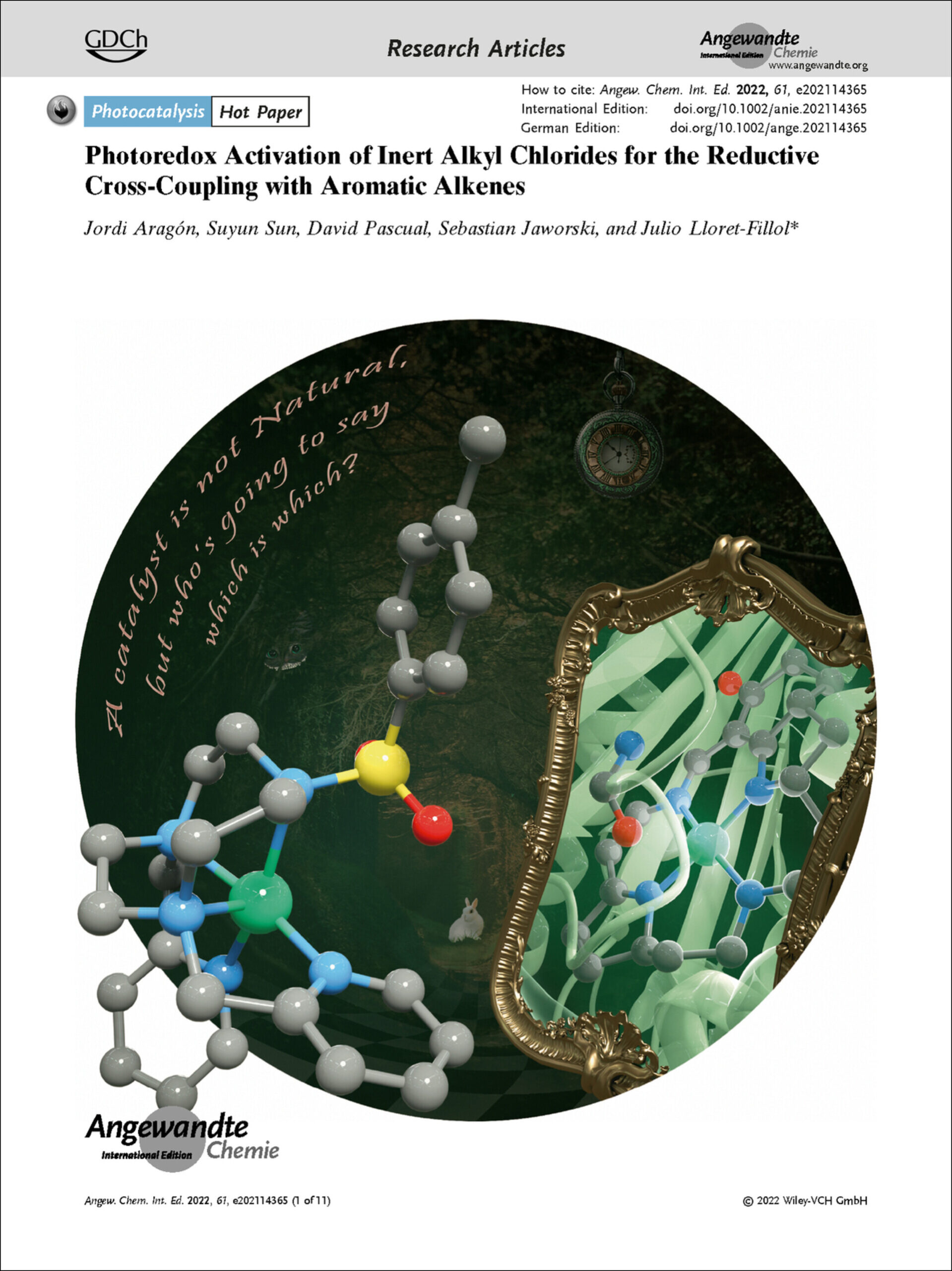
A catalyst is not Natural, but who’s going to say which is which?
This cover art draws inspiration from the classic tale of Alice in Wonderland. In this illustration, two distinct molecules are depicted gazing at each other through a mirror—one natural and the other chemically synthesized. Remarkably, their properties are so strikingly similar that they become indistinguishable. This image captures the essence of Julio Lloret-Fillol and his team’s research at the Institute of Chemical Research of Catalonia (ICIQ).
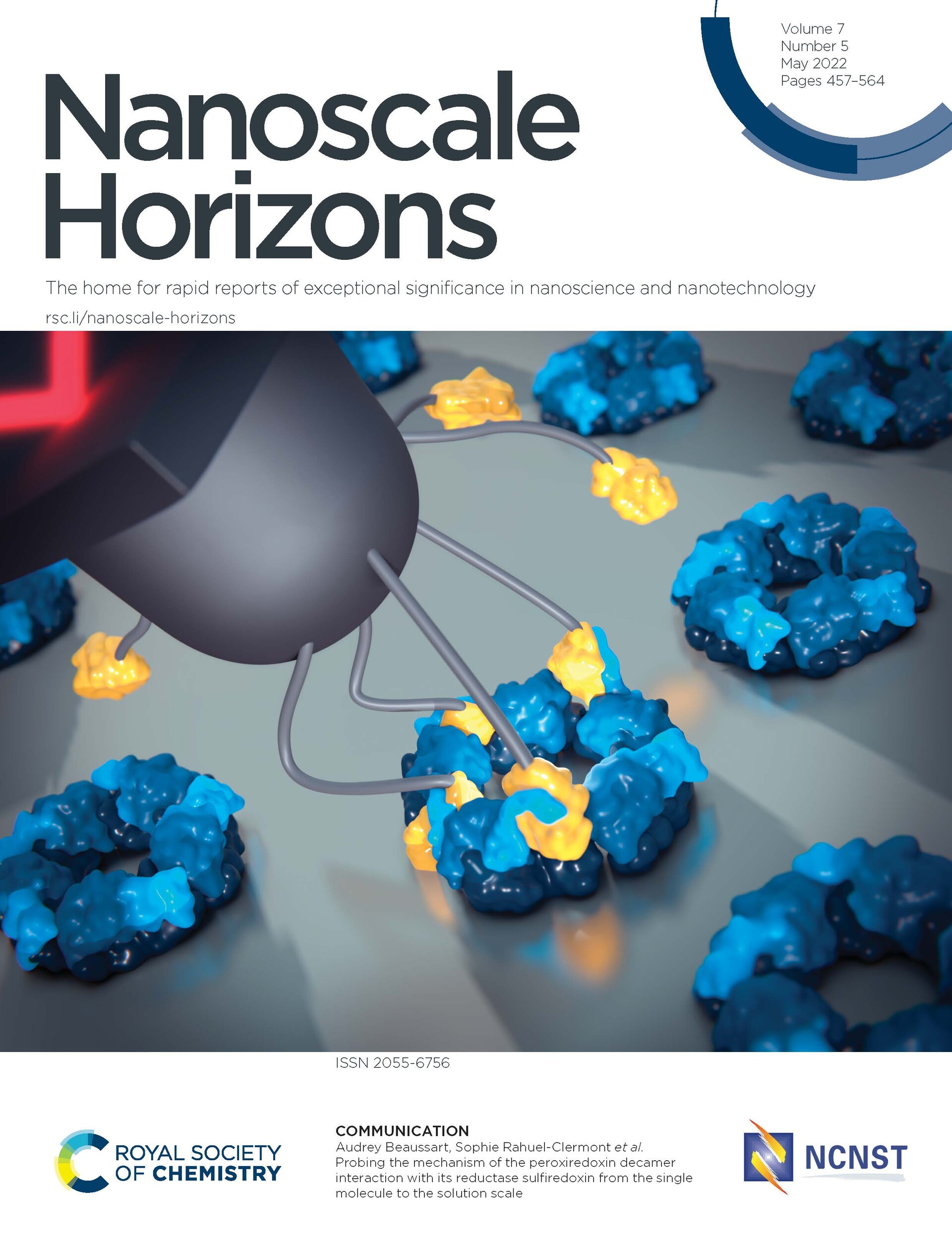
Unraveling peroxiredoxin-sulfiredoxin interaction dynamics
This illustration of Nanoscale Horizons showcases a multiscale strategy employed by Audrey Beaussart, Sophie Rahuel-Clermont, (Université de Lorraine, CNRS) and their team. Their innovative approach combines Atomic Force Microscopy (AFM), native mass spectrometry, and bulk solution techniques to investigate the intricate multivalent interaction between a decameric protein and its partner. By correlating data on affinity, kinetics, and single molecules, they gain a comprehensive understanding of this molecular interaction.
Tuning the Magnetic Anisotropy of Lanthanides on a Metal Substrate by Metal–Organic Coordination
This coverart is based on the research of Sofia Parreiras, Paolo Perna, David Écija (IMDEA Nanoscience) and coworkers. The magnetism of lanthanide-directed nanoarchitectures on surfaces can be drastically affected by small structural changes. In this paper, the authors report the effect of the coordination environment in the reorientation of the magnetic easy axis of dysprosium-directed metal-organic networks on Cu(111). They show that the magnetic anisotropy of lanthanide elements on surfaces can be tailored by specific coordinative metal–organic
Nanoscale View of Amyloid Photodynamic Damage
The image illustrates the impact of a light-activated molecule (ROS-ThT), effectively breaking the targeted amyloid fiber. In the context of biomedicine, this study holds crucial importance, as it explores new strategies for treating neurodegenerative diseases such as Alzheimer’s or Parkinson’s. The artwork was created for Cristina Flors’ research group, it holds particular significance as it forms the main focus of my thesis and marks the occasion of my first published cover, representing a significant milestone in my academic and illustrator journey.
This research is the result of a collaborative effort between IMDEA Nanoscience and Tokyo University.